
EXPLORE

Filter
Organ types
Breast
Lung
Prostate
Lymph node
Gastric
Colorectal
Multiorgan
Other filters
CE-IVD
Clinical
Research & Preclinical
H&E
IHC
CE-IVD

Aiforia® Breast Cancer Grading
Learn more

CE-IVD
Aiforia® Breast Cancer Grading
Automates breast cancer grading from H&E-stained whole-slide images (WSI), accurately identifying invasive carcinoma and ductal carcinoma in situ (DCIS). It objectively scores mitotic count, tubular formation, and nuclear pleomorphism, addressing key challenges of manual grading such as variability and time constraints, consistent with the Nottingham Grading System.
Learn more
Related materials:
Harmony on the horizon – could AI standardize breast cancer grading?
CE-IVD

Aiforia® Breast Cancer ER
Learn more

CE-IVD
Aiforia® Breast Cancer ER
Automatically detects invasive carcinoma and quantifies ER-positive and negative tumor cells from whole-slide images (WSI) or selected tissue areas, supporting consistent and objective scoring.
Learn more
CE-IVD

Aiforia® Breast Cancer PR
Learn more

CE-IVD
Aiforia® Breast Cancer PR
Automatically detects invasive carcinoma and quantifies PR-positive and negative cells from whole-slide images (WSI) or selected tissue regions, enhancing diagnostic clarity and workflow.
Learn more
CE-IVD

Aiforia® Breast Cancer HER2
Learn more

CE-IVD
Aiforia® Breast Cancer HER2
Accurately identifies invasive carcinoma and scores HER2 expression in alignment with CAP guidelines, improving diagnostic reliability and workflow efficiency.
Learn more
Related materials:
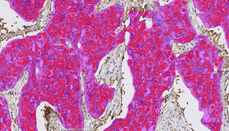
CE-IVD

Aiforia® Breast Cancer Ki67
Learn more

CE-IVD
Aiforia® Breast Cancer Ki67
Automatically detects invasive carcinoma and quantifies Ki67-positive and negative tumor cells from whole-slide images (WSI) or targeted image regions, improving diagnostic accuracy and workflow.
Learn more
CE-IVD

Aiforia® Lung Cancer PD-L1
Learn more

CE-IVD
Aiforia® Lung Cancer PD-L1
Automatically detects invasive carcinoma and delivers standardized, reproducible tumor proportion scoring (TPS), enhancing diagnostic accuracy and efficiency. It assists pathologists in overcoming the challenges of subjective and time-consuming manual PD-L1 scoring in lung cancer.
Learn more
CE-IVD

Aiforia® Prostate Cancer Biopsy
Learn more

CE-IVD
Aiforia® Prostate Cancer Biopsy
Automatically identifies adenocarcinoma, streamlines the Gleason grading process, and directly addresses interobserver variability and subtle pattern detection challenges commonly faced by pathologists.
Learn more
CE-IVD

Aiforia® Prostate Cancer PNI
Learn more

CE-IVD
Aiforia® Prostate Cancer PNI
Efficiently detects subtle perineural invasion (PNI) features, addressing the common diagnostic challenge of accurately and consistently identifying tumor involvement around nerves.
Learn more
Related materials:
CE-IVD

Aiforia® Prostate Cancer G4 Cribriform
Learn more

CE-IVD
Aiforia® Prostate Cancer G4 Cribriform
Automatically detects Gleason Grade 4 Cribriform patterns in prostate biopsy whole slide images (WSI), addressing pathologists' challenges with subjective, time-consuming visual assessments, with the aim to reduce diagnostic variability and enhance workflow efficiency.
Learn more
Related materials:

Aiforia® Prostate Cancer HG-PIN
Learn more

Aiforia® Prostate Cancer HG-PIN
Supports the detection of high-grade prostatic intraepithelial neoplasia (high-grade PIN) in whole slide images (WSI) of prostate tissue.
Learn more
CE-IVD

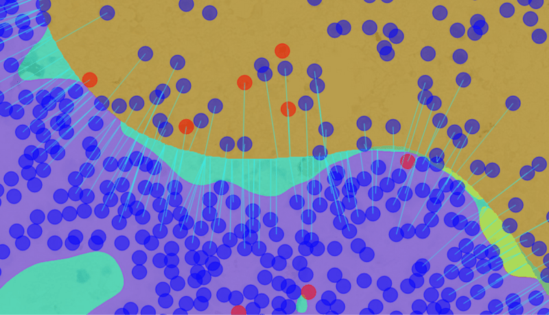
Aiforia® Lymph Node Metastasis
Learn more

CE-IVD
Aiforia® Lymph Node Metastasis
Supports the detection and quantification of metastases of breast cancer, melanoma, and colorectal cancer in lymph nodes from whole slide images.
CE-IVD

Aiforia® Gastric Cancer
Learn more

CE-IVD
Aiforia® Gastric Cancer
AI solution assisting pathologists in identifying carcinoma areas in digitized H&E-stained gastric tissue samples. It supports diagnostic decision-making while ensuring pathologists maintain full control to review or modify AI findings before final report approval.
Learn more

Helicobacter pylori (H. pylori)
Learn more

Helicobacter pylori (H. pylori)
Supports identification of Helicobacter pylori in gastric tissue samples, where bacteria may be sparse, unevenly distributed, or difficult to detect by manual review alone.

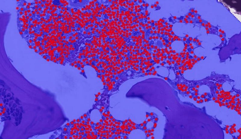
Aiforia® Colorectal Cancer QuantCRC
Learn more

Aiforia® Colorectal Cancer QuantCRC
Identifies important histological features of colorectal cancer and provides a recurrence prediction estimate useful for treatment decisions. This prognostic AI model was developed and validated in collaboration with the Mayo Clinic.
Learn more

Aiforia® Mitosis Epithelial
Learn more

Aiforia® Mitosis Epithelial
To address the challenges of time-consuming and labor-intensive mitotic counting, Aiforia® Mitosis Epithelial automates the process by spotting areas of high mitotic activity and counting the mitoses in 10 HPFs in H&E-stained whole-slide images (WSI) of epithelial tumors. AI-driven analysis and automated reporting improve efficiency and consistency in the workflow while still giving the user full control.

Aiforia® Quality Control IHC
Learn more

Aiforia® Quality Control IHC
Manual quality control of IHC-stained whole-slide images (WSI) is a time-consuming and error-prone process, often leading to delays in diagnosis. Aiforia® Quality Control IHC automates this by identifying high-quality tissue and detecting the most common artifacts. This streamlines the digital pathology workflow, saving valuable resources while ensuring high-quality images for accurate diagnostics.
Explore the case studies from Aiforia's users
Case study: enhancing mesothelioma research with AI
A research team from the University of Turin used Aiforia® Create to build an AI model for mesothelioma subtyping with reticulin stain. Read more or watch the video interview.
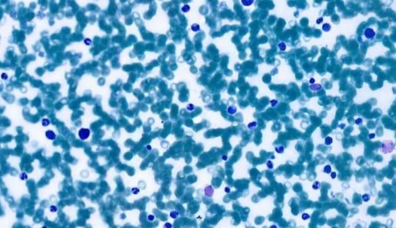
Case study: automated detection and classification of bone marrow cells
Developing a deep learning algorithm for the automated detection and classification of bone marrow aspirate smears in cytological preparations.
/Other%20medical%20areas%20JPEG/kidney_cortex_medulla_after.jpeg?width=800&height=459&name=kidney_cortex_medulla_after.jpeg)
Galileo case study: exploring AI’s potential in kidney transplantation
An Italian research team built an AI model for kidney pathology to assist on-call pathologists and renal pathologists in their routine work.

Orion pharma case study: accelerating preclinical neurotoxicity analysis with AI
Scientists at the pharmaceutical company Orion Pharma developed artificial intelligence models to automate preclinical neurotoxicity assessment.

MIT case study: advancing lung cancer research with AI
Reseachers at the Tyler Jacks Lab, MIT, created artificial intelligence models to automate tumor grading as part of their lung cancer research studies.

Massachusetts General Hospital case study: AI-assisted image analysis of neurodegenerative disease markers
Researchers from Massachusetts General Hospital used Aiforia’s AI for the analysis of histopathological markers in neurodegenerative diseases.
Faron Pharmaceuticals case study: using AI to perform spatial analysis in cancer drug development
Elisa Vuorinen at Faron Pharmaceuticals built an AI model to quantify and localize Clever-1 in the tumor microenvironment using Aiforia® Create.

Sanofi case study: Parkinson's disease research with AI
The preclinical research team studying Parkinson's disease at Sanofi created their own AI model with Aiforia® Create to automate Th+ neuron quantification.
CRL case study: AI-assisted screening of bone marrow cellularity changes
CRL Veterinary Pathologist Mark Smith describes using AI models to screen for bone marrow cellularity changes.

Experimentica case study: accelerating preclinical analysis of ocular diseases
Scientists at the CRO Experimentica describe using AI to analyze Spectral Domain Optical Coherence Tomography scans to identify neovascular lesions.