Clinical

  • Clinical Solutions
  • AI solutions portfolio

Explore Aiforia’s comprehensive portfolio of high-performance AI solutions, engineered for both clinical and preclinical applications. Aiforia’s solutions aim to enhance the speed, accuracy, and consistency of analyzing large and complex medical images, especially in pathology.

Learn more

Research

  • Research solutions
  • Aiforia® Create
  • AI solutions portfolio

Aiforia’s research solutions offer fully automated study-centric workflows and pathology image analysis applications that cater to all research areas. Empowered by robust AI capabilities, pathologists and scientists can make discoveries while saving time by automating repetitive tasks – also within the framework of GLP. Aiforia’s solutions can be integrated with any existing laboratory infrastructure to enjoy the full benefits of a digitized workflow.

Learn more

Aiforia® Create is the most versatile tool for developing deep learning AI models for image analysis in digital pathology. Its cloud-based, collaborative working environment allows multiple users to work together in real time, anywhere in the world. Praised for its intuitive user interface, it allows users a fast start, even without any prior AI experience.Learn more >

Learn more

Explore Aiforia’s comprehensive portfolio of high-performance AI solutions, engineered for both clinical and preclinical applications. Aiforia’s solutions aim to enhance the speed, accuracy, and consistency of analyzing large and complex medical images, especially in pathology.

Learn more

AI development tool

  • Aiforia® Create

Aiforia® Create is the most versatile tool for developing deep learning AI models for image analysis in digital pathology. Its cloud-based, collaborative working environment allows multiple users to work together in real time, anywhere in the world. Praised for its intuitive user interface, it allows users a fast start, even without any prior AI experience.

Learn more

Resources

  • Publications
  • Resource Library
  • Press releases

Explore some of our latest publications showcasing the discoveries enabled by the Aiforia® Platform.

Learn more

Aiforia's insights and resources for AI in pathology. View some of our content pieces from the world of AI.

Learn more

Stay updated with Aiforia’s recent announcements. Explore our latest news, updates and announcements.

Learn more

Company

  • About us
  • Investors
  • Partners
  • Media
  • Events
  • Careers
  • Quality and security

Find out more about shareholder matters, investment discussions, and financial-related information.

Learn more

Aiforia's mission is to transform pathology image analysis with AI, enabling better care for each patient. To succeed, we need our trusted partners. Explore Aiforia’s partner network.

Learn more

Experience our AI solutions firsthand. Explore the list of conferences where you can connect with Aiforia’s team live and book a time to discover the future of AI-powered image analysis.

Learn more

Our mission is to transform pathology image analysis with AI, enabling better care for each patient. Find out more about job openings, thesis opportunities, or submitting applications.

Learn more

Aiforia Technologies holds information security to the highest standard. We hold several, globally relevant quality and security certifications, so you can be sure your sensitive information is safe.

Learn more

Research

Research solutions

Aiforia® Create

AI solutions portfolio

AI development tool

Aiforia® Create

Resources

Publications

Resource Library

Press releases

Aiforia® Lung Cancer Suite

AI for lung cancer diagnostics

Overcome the challenges of subjective and time-consuming manual PD-L1 scoring in lung cancer – Aiforia® Lung Cancer Suite can reduce errors and help standardize scoring by providing AI-driven PD-L1 assessments that are objective and reproducible. 

Request a demo
Clinical Image 3
PD-L1_monitor_right angle

Enhance diagnostic accuracy with AI-driven PD-L1 analysis

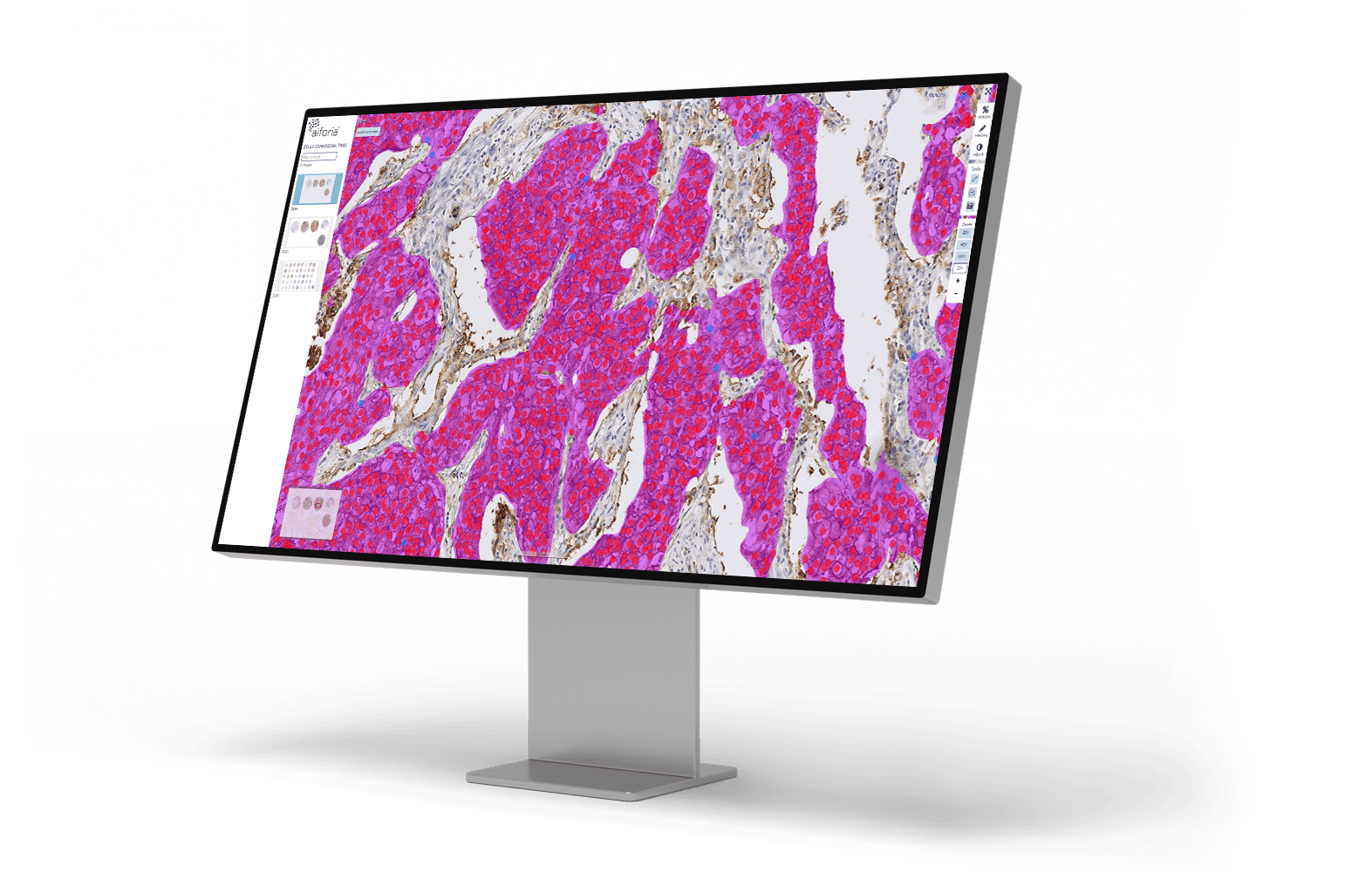
PD-L1 is a critical biomarker to guide immunotherapies in lung cancer care, and even minor differences in the analysis of PD-L1 expression can drastically alter treatment plans. Currently, PD-L1 is scored by a pathologist by visual estimation (“eyeballing”), which provides high accuracy in clear-cut cases. Still, it introduces variability of results in the middle ranges of PD-L1 expression with potentially drastic effects in the patient care: Being off by even 5 or 10 percentage points can shift a patient’s treatment regime entirely; under-calling can deny patients effective immunotherapy, while over-calling can lead to unnecessary treatment.

AI assistance brings consistency and efficiency to the PD-L1 scoring: AI algorithms apply the same criteria to every slide, aiming for objective reproducibility. AI can examine every tumor cell across the whole slide (hundreds or thousands), and once trained, AI can score PD-L1 in minutes, accelerating turnaround and freeing pathologists from tedious counting. 

Lung Cancer Suite: AI solution

Aiforia® Lung Cancer PD-L1
Lung_PDL1_20x_1_AImasks_cropped

Automatically detects invasive carcinoma and delivers standardized, reproducible tumor proportion scoring (TPS), enhancing diagnostic accuracy and efficiency. It assists pathologists in overcoming the challenges of subjective and time-consuming manual PD-L1 scoring in lung cancer.

Clinical performance highlights
  • Excellent diagnostic accuracy with high correlation to expert visual assessments
  • Enhanced consistency through improved pathologist concordance (Fleiss’ Kappa ~0.87)
  • Significantly streamlined workflow, reducing reporting time by up to 46%
  • Reliable and standardized scoring at critical clinical thresholds (e.g., 1% and 50%)


CE-IVD marked for diagnostic use in EU and EEA countries, and for Research Use Only (RUO) and Performance Studies Only (PSO) in all other market areas

Lung Cancer Suite: AI solution

Aiforia® Lung Cancer PD-L1

Lung_PDL1_20x_1_AImasks_cropped

Automatically detects invasive carcinoma and delivers standardized, reproducible tumor proportion scoring (TPS), enhancing diagnostic accuracy and efficiency. It assists pathologists in overcoming the challenges of subjective and time-consuming manual PD-L1 scoring in lung cancer.

Clinical performance highlights
  • Excellent diagnostic accuracy with high correlation to expert visual assessments
  • Enhanced consistency through improved pathologist concordance (Fleiss’ Kappa ~0.87)
  • Significantly streamlined workflow, reducing reporting time by up to 46%
  • Reliable and standardized scoring at critical clinical thresholds (e.g., 1% and 50%)


CE-IVD marked for diagnostic use in EU and EEA countries, and for Research Use Only (RUO) and Performance Studies Only (PSO) in all other market areas

Why Aiforia® Clinical Suites?

Industry_CRO

Precise, transparent AI results

Aiforia takes accuracy beyond standard AI heatmaps by clearly marking the exact tumor areas with detailed visual overlays directly on the digital slides, enabling easy visual confirmation at any magnification level. This approach empowers pathologists with both visual transparency and exact quantitative data, such as accurate tumor area measurements and cell counts, delivering actionable insights.

Products_Clinical

Comprehensive case-level review and reporting

Aiforia® Clinical Suites conveniently combine results from multiple AI models, WSIs, and stainings into a case-level view and report for first-read diagnostic support.

ic_save_time_pink

Speeds up each step in your workflow

The AI results are populated directly into the reporting panel. The user can utilize advanced features like case list triaging, pre-ordering of extra stains, and flagging of non-representative samples for a faster workflow.

ic_our_ai-1_pink

Increases the reliability of PD-L1 analysis

Aiforia® Lung Cancer PD-L1 provides standardized scoring, with high accuracy, increasing the reliability of PD-L1 analysis.

Case studies, expert opinions, and more

Resources for lung cancer pathology

PD-L1_stock 1_resource_carousel_banner-2

WEBINAR

PD-L1 on the edge: cut-offs, cognitive bias & the case for AI in lung cancer pathology

This webinar takes you inside the real-world diagnostic pressure points of PD-L1 TPS testing in NSCLC.

Watch recording >

Lung_PDL1_carousel-size

CASE STUDY

Southampton case study: AI-driven analysis of PD-L1 lung cancer

University Hospital Southampton is implementing Aiforia’s clinical PD-L1 lung cancer AI solution to assess PD-L1 staining in lung cancer specimens.

Read more >

case-study-dr-boland-et-al-lung-adenocarcinoma

CASE STUDY

Developing an AI model to determine invasion in pulmonary adenocarcinoma

Dr. Jennifer M. Boland et al. from the Mayo Clinic successfully developed an AI model to determine the invasion in pulmonary adenocarcinoma.

Read more >

dr-smith-case-study-advanced-fibrosis-carousel

CASE STUDY

Differentiating interstitial lung disease subtypes using AI-based image analysis

Dr. Maxwell L. Smith from Mayo Clinic developed two AI models to detect and quantify histopathologic features in interstitial lung disease (ILD) with the goal of using AI to help differentiate ILD subtypes.

Read the case >

PD-L1-aiforward-project-carousel

CASE STUDY

Automating the quantification of PD-L1 in lung cancer

Liesbeth Hondelink, MSc student at Leiden University Medical Centre, describes using AI to automate the quantification of PD-L1 in lung cancer studies.

Read more >

PD-L1-after-carousel

CASE STUDY

Validation of PD-L1 AI model in six labs in the Netherlands

Labs in the Netherlands are joining together to launch an AI model validation study for PD-L1 scoring with a goal of multi-source domain adaptation.

Read more >


Seeing is believing

Request a demo

Discover the power of AI for image analysis

Find out how to enhance your image analysis work in diagnostic pathology, preclinical studies, or medical research. The demo will be tailored based on your interests.

The demo will help you understand:

  • How AI-assisted image analysis can increase efficiency, precision, and consistency in the pathology workflow.
  • The limitless possibilities Aiforia® Platform offers – both for research and clinical diagnostics – and the suitable use cases for your needs.

We can either demonstrate Aiforia’s image analysis solutions on your own images or any of our application examples (i.e., neuron quantification, automated tumor grading, NASH analysis, etc.).

Fill in the form, and one of our experts will contact you shortly to schedule the time.