Aiforia® Gastric Suite

AI for gastric diagnostics

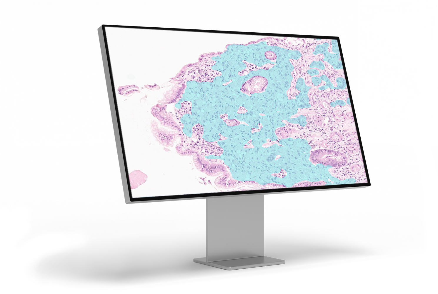
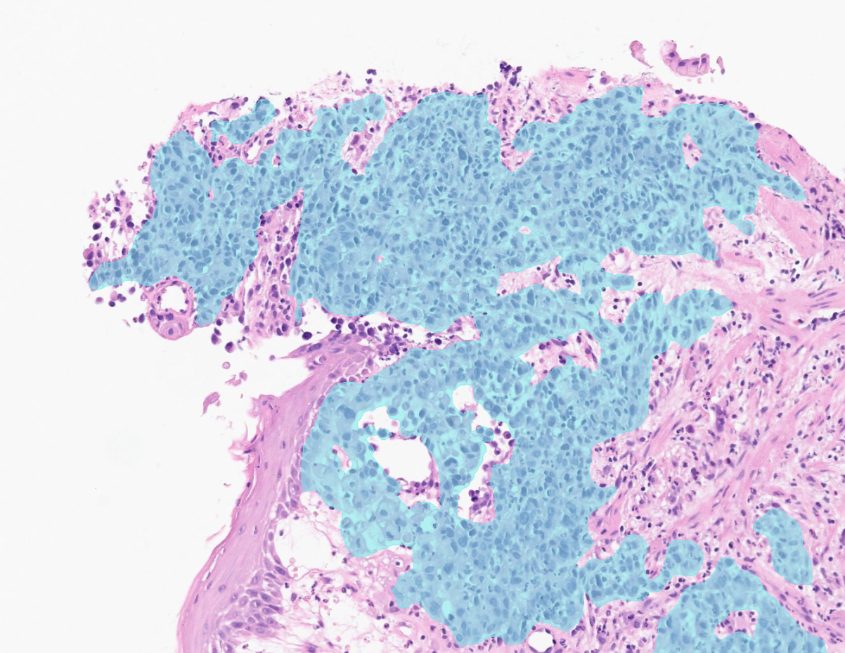
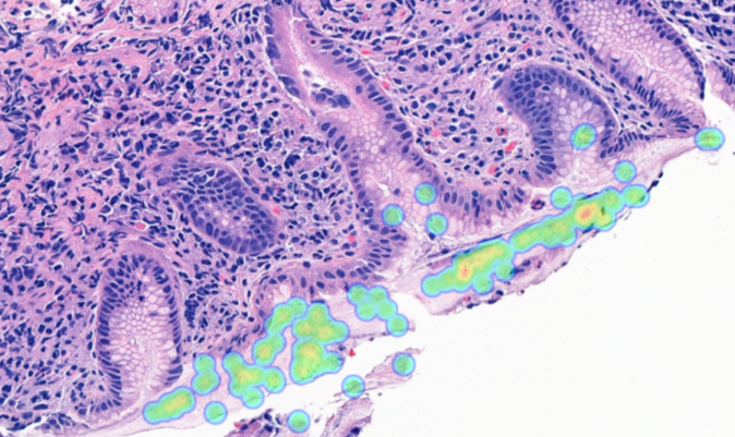
Manual screening for gastric carcinoma and Helicobacter pylori (H. pylori) is time-consuming and challenging. Subtle tumor foci and low bacterial density are subject to interobserver variability and workload pressure.

Aiforia® Gastric Suite uses AI to analyze digitized whole slide images to address these challenges. By highlighting suspicious areas directly on the slide, it enables efficient review while ensuring the pathologist remains in full control of interpretation and reporting.

Contact us
Gastric Cancer_clinical_stock 1-1
Gastric cancer_monitor left angle_1-1

Enhance diagnostic confidence with AI-assisted gastric pathology

Subtle, fragmented, or poorly cohesive gastric carcinoma can be difficult to detect, even for experienced pathologists. Tumor foci identification requires meticulous manual screening, where even a minor oversight may significantly impact patient outcomes.

Aiforia® Gastric Cancer AI model mitigates these risks by providing a systematic, AI-driven analysis of whole slide images. By flagging suspicious regions and applying consistent diagnosis criteria, it reduces interobserver variability and ensures high-risk areas are not overlooked. AI results are automatically visualized on the digital slide and populated on the case-level report, enabling faster review.

Gastric Suite: AI models

Aiforia® Gastric Cancer
Helicobacter pylori (H. pylori)
Gastric cancer model-1

Aiforia® Gastric Cancer is a CE-IVD marked AI solution assisting pathologists in identifying carcinoma areas in digitized H&E-stained gastric tissue samples. It supports diagnostic decision-making while ensuring pathologists maintain full control to review or modify AI findings before final report approval. Built on Aiforia’s Foundation Engine technology, it provides robust, high-performance results across real-world variations in sample quality, staining, and scanning.

Clinical performance highlights
  • Designed for high sensitivity - supports the detection of suspicious regions, no matter the size or location of the lesion
  • Supports consistent interpretation across different readers
  • Integrates seamlessly into routine digital pathology workflows


CE-IVD marked for diagnostic use in EU and EEA countries (IVDR) and for Research Use Only (RUO) and Performance Studies Only (PSO) in all other market areas.

Gastric_H-Pylori

The H. pylori AI model supports identification of Helicobacter pylori in gastric tissue samples, where bacteria may be sparse, unevenly distributed, or difficult to detect by manual review alone.

By highlighting areas of interest directly on the whole slide image, the model helps guide the pathologist's attention and supports efficient verification.


H. pylori AI model is currently for Research Use Only (RUO) and for Performance Studies Only (PSO) in all market areas.


Gastric Suite: AI models

Aiforia® Gastric Cancer

Gastric cancer model-1

Aiforia® Gastric Cancer is a CE-IVD marked AI solution assisting pathologists in identifying carcinoma areas in digitized H&E-stained gastric tissue samples. It supports diagnostic decision-making while ensuring pathologists maintain full control to review or modify AI findings before final report approval. Built on Aiforia’s Foundation Engine technology, it provides robust, high-performance results across real-world variations in sample quality, staining, and scanning.

Clinical performance highlights
  • Designed for high sensitivity - supports the detection of suspicious regions, no matter the size or location of the lesion
  • Supports consistent interpretation across different readers
  • Integrates seamlessly into routine digital pathology workflows


CE-IVD marked for diagnostic use in EU and EEA countries (IVDR) and for Research Use Only (RUO) and Performance Studies Only (PSO) in all other market areas.

Helicobacter pylori (H. pylori)

Gastric_H-Pylori

The H. pylori AI model supports identification of Helicobacter pylori in gastric tissue samples, where bacteria may be sparse, unevenly distributed, or difficult to detect by manual review alone.

By highlighting areas of interest directly on the whole slide image, the model helps guide the pathologist's attention and supports efficient verification.


H. pylori AI model is currently for Research Use Only (RUO) and for Performance Studies Only (PSO) in all market areas.


Why Aiforia® Clinical Suites?

Industry_CRO

Precise, transparent AI results

Aiforia takes accuracy beyond standard AI heatmaps by clearly marking the exact tumor areas with detailed visual overlays directly on the digital slides, enabling easy visual confirmation at any magnification level. This approach empowers pathologists with both visual transparency and exact quantitative data, such as accurate tumor area measurements and cell counts, delivering actionable insights.

Products_Clinical

Comprehensive case-level review and reporting

Aiforia® Clinical Suites conveniently combine results from multiple AI models, WSIs, and stainings into a case-level view and report for first-read diagnostic support.

ic_save_time_pink

Speeds up each step in your workflow

The AI results are populated directly into the reporting panel. The user can utilize advanced features like case list triaging, pre-ordering of extra stains, and flagging of non-representative samples for a faster workflow.

ic_discover_pink

Built for difficult and easily missed gastric cancer cases

Gastric cancer often presents with small, scattered, or poorly cohesive tumor cells that can be challenging to detect, particularly in biopsy material. Aiforia® Gastric Cancer is designed to support detection across major histological patterns of gastric carcinoma, helping reduce the risk of missed findings in routine practice.

Case studies, expert opinions, and more

Clinical implementation stories from Aiforia’s customers

Memorial Hospital_carousel_size-1

CASE STUDY

Memorial Pathology case study: implementing AI in a private hospital setting

Memorial Pathology implemented Aiforia® Clinical Suites for breast, prostate, and PD-L1 lung to gain efficiency and accuracy in the routine workflow.

Read more >

APHP press release_blog banner-2

BLOG POST

Guest blog: evolution of artificial intelligence in diagnostic pathology

Dr. Catherine Guettier reflects on the implementation of AI in pathology workflows across French hospitals.

Read more >

Blog post Democratizing AI-1

BLOG POST

How did the Mayo Clinic choose a vendor for AI in pathology?

What to consider when choosing a vendor for AI in pathology? Learn how the Mayo Clinic evaluated platform providers based on six key criteria.

Read the case >

Digital pathology deployment in Veneto 8 steps to success_blog banner-1-1-4

BLOG POST

Digital pathology deployment in Veneto: 8 steps to success

What is required for pathology laboratories to go fully digital? Learn how the broadest public healthcare provider in the Veneto region did it.

Read more >


CONTACT US

Discover the power of AI for image analysis

Contact us to learn more about how Aiforia’s AI can enhance your diagnostic process and tailor it to your laboratory’s unique needs.

We will help you understand: 

  • How AI-assisted image analysis can increase efficiency, precision, and consistency in the pathology workflow.
  • The limitless possibilities the Aiforia® Platform offers and the suitable solutions for your needs.

Fill in the form, and one of our experts will contact you shortly.