Clinical

  • Clinical Solutions
  • AI solutions portfolio

Explore Aiforia’s comprehensive portfolio of high-performance AI solutions, engineered for both clinical and preclinical applications. Aiforia’s solutions aim to enhance the speed, accuracy, and consistency of analyzing large and complex medical images, especially in pathology.

Learn more

Research

  • Research solutions
  • Aiforia® Create
  • AI solutions portfolio

Aiforia’s research solutions offer fully automated study-centric workflows and pathology image analysis applications that cater to all research areas. Empowered by robust AI capabilities, pathologists and scientists can make discoveries while saving time by automating repetitive tasks – also within the framework of GLP. Aiforia’s solutions can be integrated with any existing laboratory infrastructure to enjoy the full benefits of a digitized workflow.

Learn more

Aiforia® Create is the most versatile tool for developing deep learning AI models for image analysis in digital pathology. Its cloud-based, collaborative working environment allows multiple users to work together in real time, anywhere in the world. Praised for its intuitive user interface, it allows users a fast start, even without any prior AI experience.Learn more >

Learn more

Explore Aiforia’s comprehensive portfolio of high-performance AI solutions, engineered for both clinical and preclinical applications. Aiforia’s solutions aim to enhance the speed, accuracy, and consistency of analyzing large and complex medical images, especially in pathology.

Learn more

AI development tool

  • Aiforia® Create

Aiforia® Create is the most versatile tool for developing deep learning AI models for image analysis in digital pathology. Its cloud-based, collaborative working environment allows multiple users to work together in real time, anywhere in the world. Praised for its intuitive user interface, it allows users a fast start, even without any prior AI experience.

Learn more

Resources

  • Publications
  • Resource Library
  • News room

Explore some of our latest publications showcasing the discoveries enabled by the Aiforia® Platform.

Learn more

Aiforia's insights and resources for AI in pathology. View some of our content pieces from the world of AI.

Learn more

Stay updated with Aiforia’s recent announcements. Explore our latest news, updates and announcements.

Learn more

Company

  • About us
  • Investors
  • Partners
  • Media
  • Events
  • Careers
  • Quality and security

Find out more about shareholder matters, investment discussions, and financial-related information.

Learn more

Aiforia's mission is to transform pathology image analysis with AI, enabling better care for each patient. To succeed, we need our trusted partners. Explore Aiforia’s partner network.

Learn more

Experience our AI solutions firsthand. Explore the list of conferences where you can connect with Aiforia’s team live and book a time to discover the future of AI-powered image analysis.

Learn more

Our mission is to transform pathology image analysis with AI, enabling better care for each patient. Find out more about job openings, thesis opportunities, or submitting applications.

Learn more

Aiforia Technologies holds information security to the highest standard. We hold several, globally relevant quality and security certifications, so you can be sure your sensitive information is safe.

Learn more

Research

Research solutions

Aiforia® Create

AI solutions portfolio

AI development tool

Aiforia® Create

Resources

Publications

Resource Library

News room

Aiforia® Colon Suite

AI for colon diagnostics

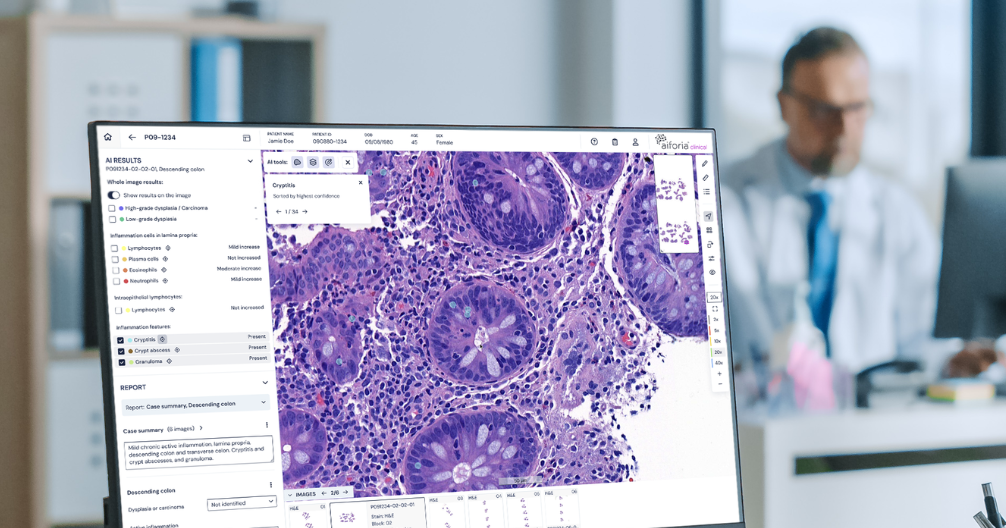
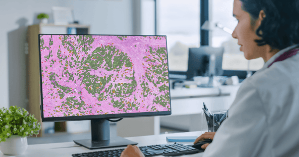
Aiforia® Colon Suite leverages AI-driven image analysis on digitized tissue samples, automating triaging and highlighting key findings directly on the slide. It is designed to streamline the digital pathology workflows and provide diagnostic support while ensuring the pathologist remains in full control of interpretation and reporting.

Request a demo
Colonoscopy_stock image 1

Colon Suite: AI models

Aiforia® Colonoscopy
Aiforia® Lymph Node Metastasis
Aiforia® Colorectal Cancer QuantCRC
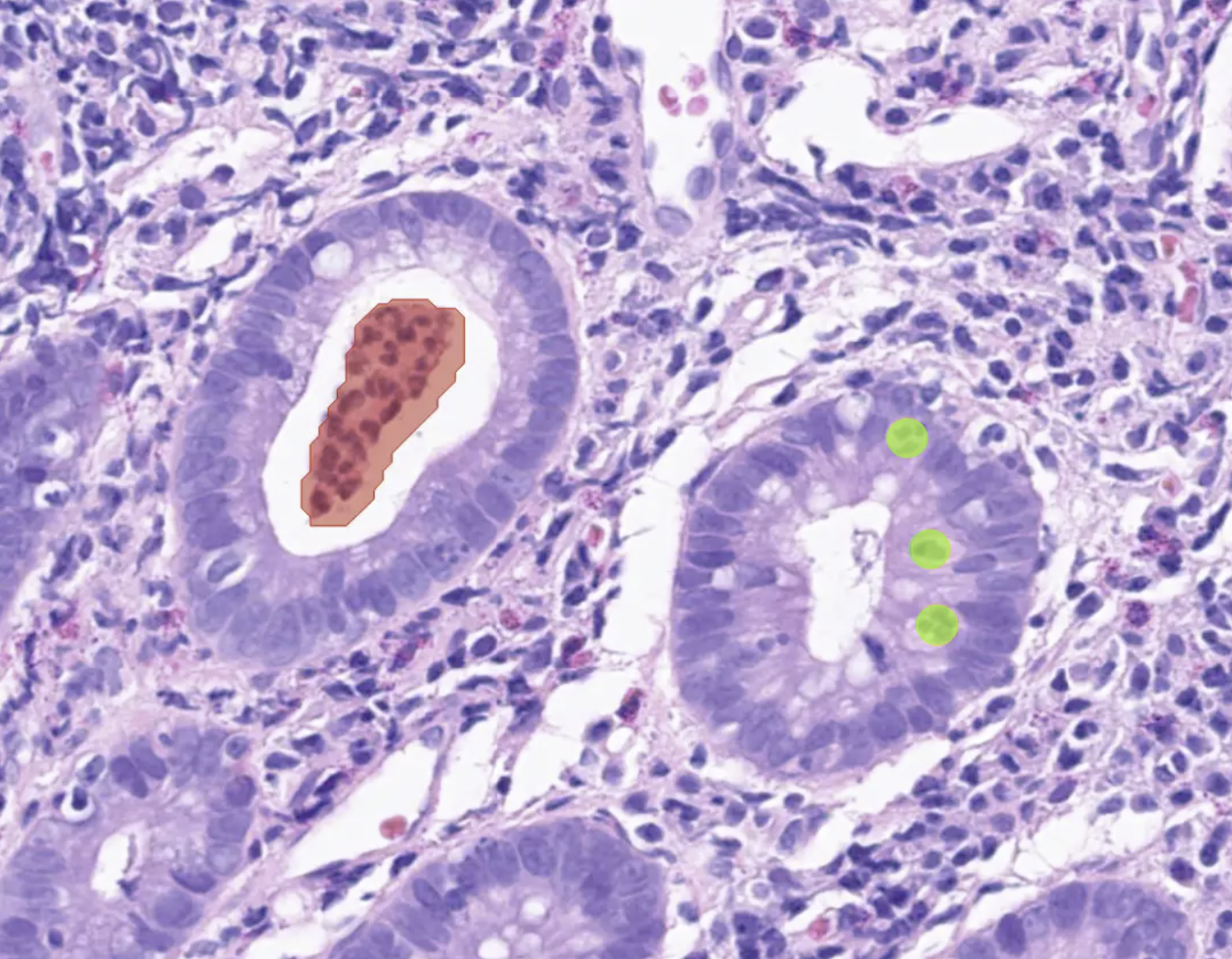
Colonoscopy_abscess and cryptitis_greenobjects

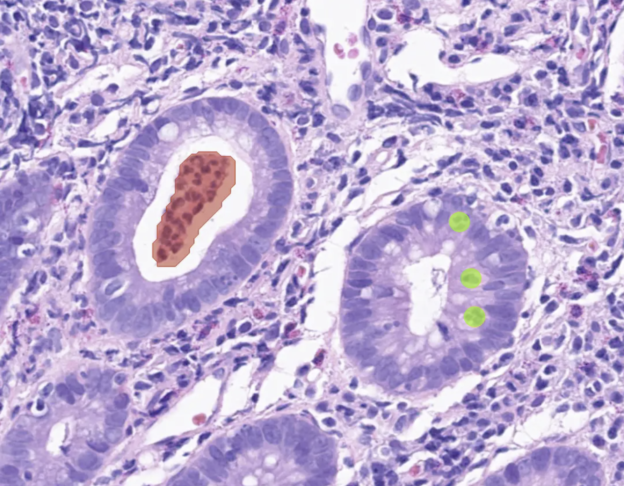
Designed to streamline the diagnostic workflow for digitized H&E-stained colonoscopy samples. By automating sample triaging and identifying a broad spectrum of key pathologies, such as carcinoma, dysplasia, and active and chronic inflammation, it aims to empower pathologists with enhanced diagnostic support.

> Contact us for availability in your territory


Aiforia® Colonoscopy is currently for Research Use Only (RUO) and for Performance Studies Only (PSO) in all market areas.

aiforia-lymph-node-metastasis-breast

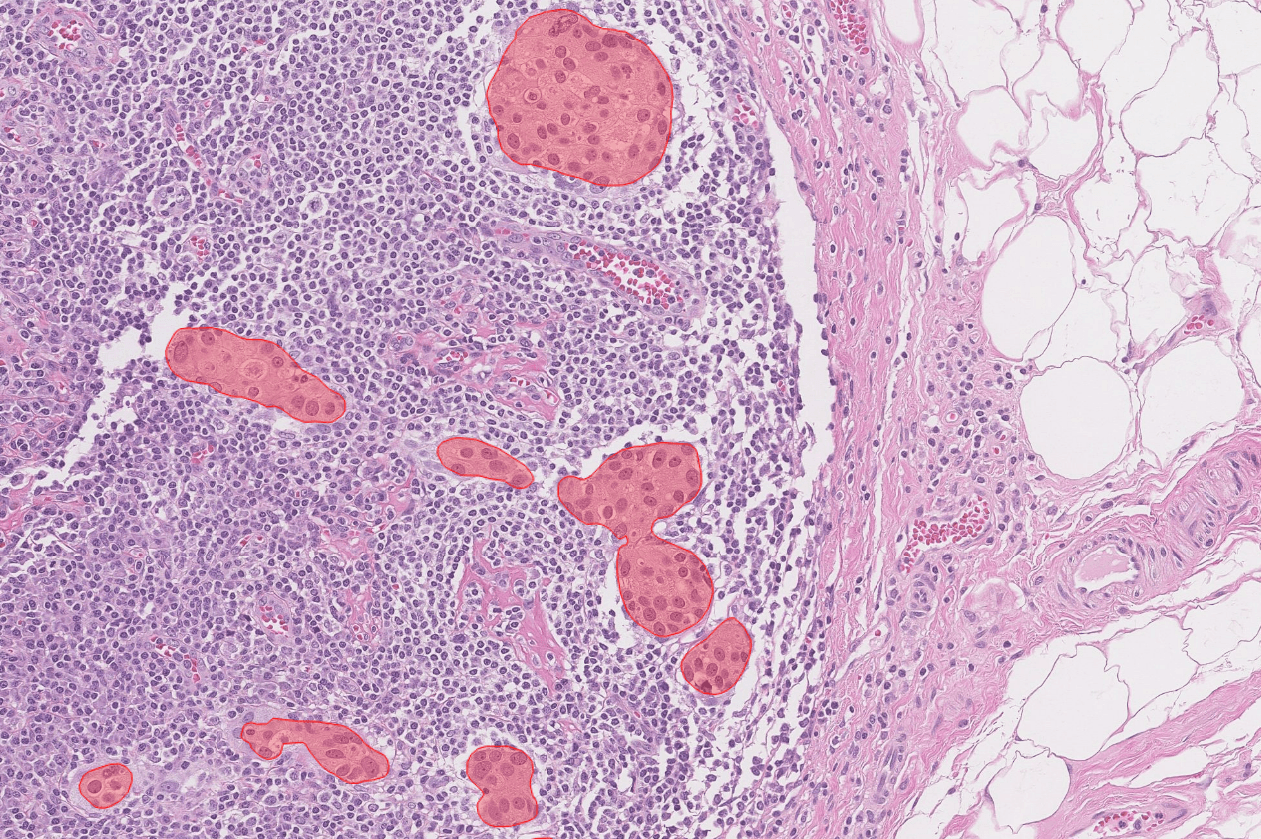
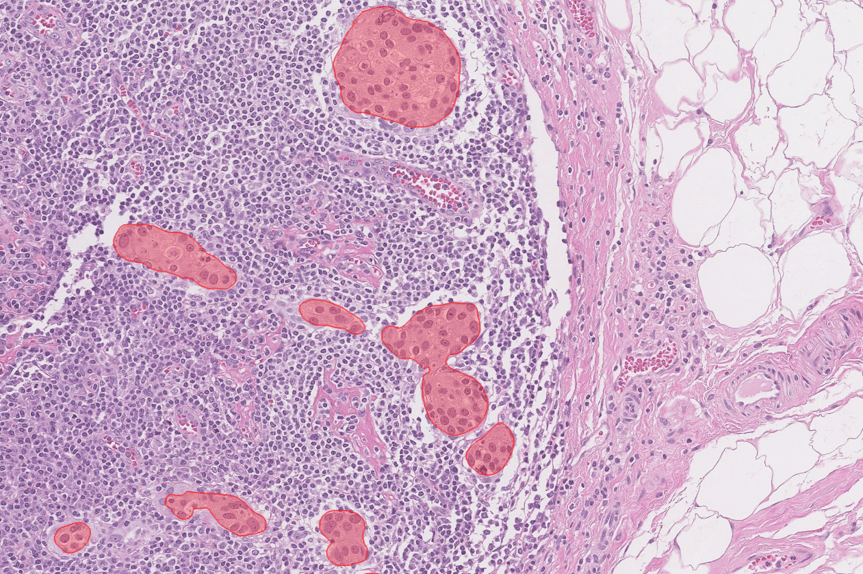
Supports the detection and quantification of metastases of breast cancer, melanoma, and colorectal cancer in lymph nodes from whole slide images.

> Contact us for more details


CE-IVD marked for diagnostic use in EU and EEA countries, and for Research Use Only (RUO) and Performance Studies Only (PSO) in all other market areas.

QuantCRC_image 1-1

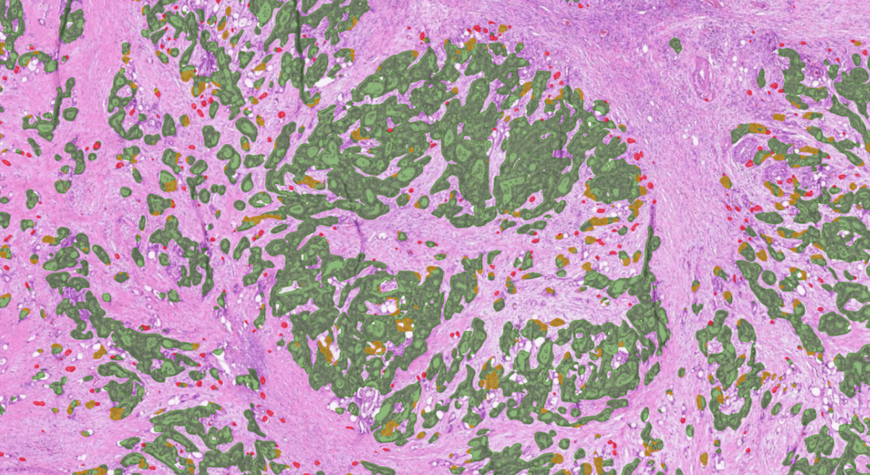
This prognostic AI model provides a recurrence-prediction estimate to support treatment decisions. The estimate is based on the identification of important histological features of colorectal cancer. The AI model was developed and validated in collaboration with the Mayo Clinic.

> Learn more


Aiforia® Colorectal Cancer QuantCRC is currently for Research Use Only (RUO) and for Performance Studies Only (PSO) in all market areas.


Colon Suite: AI models

Aiforia® Colonoscopy

Colonoscopy_abscess and cryptitis_greenobjects

Designed to streamline the diagnostic workflow for digitized H&E-stained colonoscopy samples. By automating sample triaging and identifying a broad spectrum of key pathologies, such as carcinoma, dysplasia, and active and chronic inflammation, it aims to empower pathologists with enhanced diagnostic support.

> Contact us for availability in your territory


Aiforia® Colonoscopy is currently for Research Use Only (RUO) and for Performance Studies Only (PSO) in all market areas.

Aiforia® Lymph Node Metastasis

aiforia-lymph-node-metastasis-breast

Supports the detection and quantification of metastases of breast cancer, melanoma, and colorectal cancer in lymph nodes from whole slide images.

> Contact us for more details


CE-IVD marked for diagnostic use in EU and EEA countries, and for Research Use Only (RUO) and Performance Studies Only (PSO) in all other market areas.

Aiforia® Colorectal Cancer QuantCRC

QuantCRC_image 1-1

This prognostic AI model provides a recurrence-prediction estimate to support treatment decisions. The estimate is based on the identification of important histological features of colorectal cancer. The AI model was developed and validated in collaboration with the Mayo Clinic.

> Learn more


Aiforia® Colorectal Cancer QuantCRC is currently for Research Use Only (RUO) and for Performance Studies Only (PSO) in all market areas.


Why Aiforia® Clinical Suites?

Industry_CRO

Precise, transparent AI results

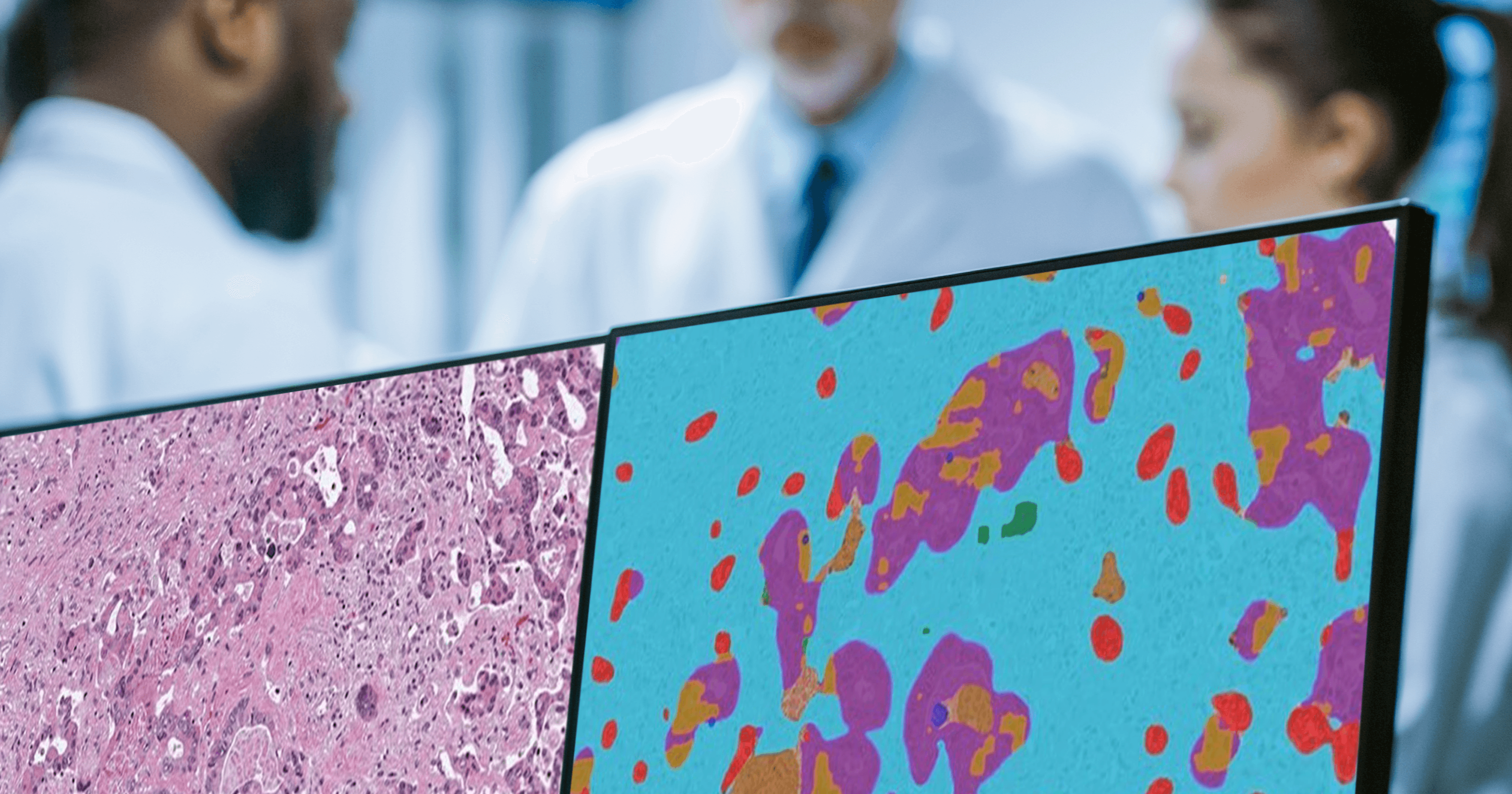
Aiforia takes accuracy beyond standard AI heatmaps by clearly marking the exact tumor areas with detailed visual overlays directly on the digital slides, enabling easy visual confirmation at any magnification level. This approach empowers pathologists with both visual transparency and exact quantitative data, such as accurate tumor area measurements and cell counts, delivering actionable insights.

Products_Clinical

Comprehensive case-level review and reporting

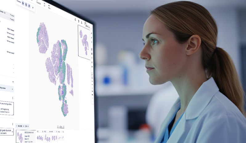
Aiforia® Clinical Suites conveniently combine results from multiple AI models, WSIs, and stainings into a case-level view and report for first-read diagnostic support.

ic_save_time_pink

Speeds up each step in your workflow

The AI results are populated directly into the reporting panel. The user can utilize advanced features like case list triaging, pre-ordering of extra stains, and flagging of non-representative samples for a faster workflow.

ic_discover_pink

Built for difficult and easily missed gastric cancer cases

Gastric cancer often presents with small, scattered, or poorly cohesive tumor cells that can be challenging to detect, particularly in biopsy material. Aiforia® Gastric Cancer is designed to support detection across major histological patterns of gastric carcinoma, helping reduce the risk of missed findings in routine practice.

Case studies, expert opinions, and more

Clinical implementation stories from Aiforia’s customers

Rising colonoscopy biopsy volumes are outpacing pathologists_blog banner-1-1

BLOG POST

Rising colonoscopy biopsy volumes are outpacing pathologists

Rising colonoscopy sample volumes are outpacing the pathology workforce. Discover how AI-assisted digital pathology helps digest the GI biopsy bottleneck.

Read more >

www-aiforia-comhubfsblog imagescolorectal_stock 3

CASE STUDY

Case study: creating a prognostic AI model for colorectal cancer

Dr. Rish Pai from the Mayo Clinic developed an AI model to assist oncologists in deciding which CRC patients should receive chemotherapy and for how long.

Read more >

Blog post Democratizing AI-1

WEBINAR

Using prognostic AI models in pathology: case colorectal cancer

Discover how artificial intelligence can aid oncologists and pathologists working in the gastrointestinal field. This webinar showcases an AI model, developed by the Mayo Clinic and Aiforia, that identifies important histological features of colorectal cancer and provides a recurrence prediction estimate useful for treatment decisions.

Watch here >

Aiforias AI video interview_Unibern_thumbnail-1

VIDEO

Industry-academia collaboration led to a CE-IVD marked AI solution: Aiforia and the University of Bern

Aiforia and the University of Bern developed a CE-IVD marked AI model for detecting lymph node metastasis from breast, colorectal, and melanoma cases. This is an excellent proof of how collaboration between academia and industry can result in solutions that will improve healthcare.

Watch here >


Seeing is believing

Request a demo

Discover the power of AI for image analysis

Find out how to enhance your image analysis work in diagnostic pathology, preclinical studies, or medical research. The demo will be tailored based on your interests.

The demo will help you understand:

  • How AI-assisted image analysis can increase efficiency, precision, and consistency in the pathology workflow.
  • The limitless possibilities Aiforia® Platform offers – both for research and clinical diagnostics – and the suitable use cases for your needs.

We can either demonstrate Aiforia’s image analysis solutions on your own images or any of our application examples (i.e., neuron quantification, automated tumor grading, NASH analysis, etc.).

Fill in the form, and one of our experts will contact you shortly to schedule the time.