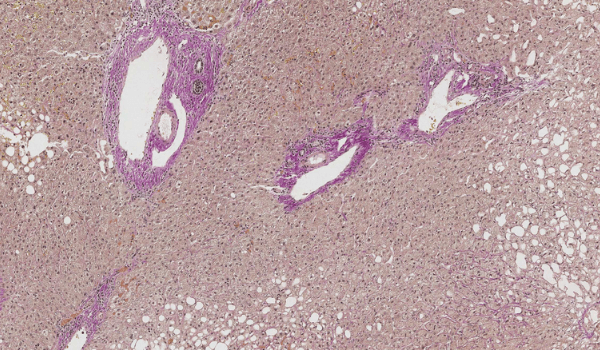
AI in liver pathology
Expand discovery with AI-based tools
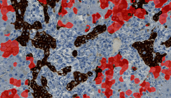
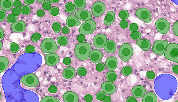
Aiforia’s solutions are created for pathologists and healthcare professionals to empower their image analysis with deep learning artificial intelligence and high-speed, cloud-based tools.
You can automate, standardize, and accelerate image analysis applications for segmentation, quantification, regression model creation, measurement of spatial and morphological metrics, analysis of serial sample sections, and so much more.
Request a demo