Introduction
PEDEGO, a research unit at the University of Oulu, covers a wide range of topics primarily around pediatrics, obstetrics, and gynecology. The metabolic health group within this unit has several ongoing clinical trials related to reproductive and metabolic health in women, including the function of the endometrium in women with polycystic ovary syndrome (PCOS). PCOS affects 10-18% of fertile aged women and many present with complications such as metabolic syndrome and infertility. It is also highly linked to obesity, with diabetes and hypertension as common comorbidities.
The inner lining of the uterus, the endometrium, is a hormone responsive tissue and consists of epithelium tightly connected with underlying stromal cells, allowing for a strong relationship between the layers. Women with PCOS present with an altered endometrial function related to steroid hormones or metabolic issues contributing to infertility. We interviewed Marika Kangasmiemi, MD and PhD student at the University of Oulu, on her experience with Aiforia Create to study the endometrium in different phases of the menstrual cycle.
Interview with Dr. Marika Kangasniemi
Tell us a bit about the project or research work you used Aiforia’s software for:
Dr. Kangasniemi: Endometrial samples from 2 different patient groups and from different phases of the menstrual cycle were analyzed so that endometrial leukocytes and proliferative cells were counted, separately in epithelium and stroma.
What is unique about the project?
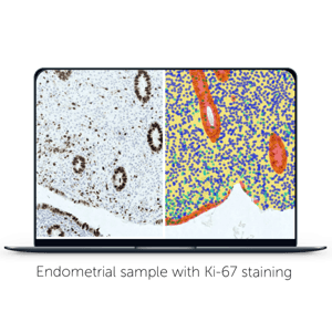
Endometrial tissue is dynamic, and the amount of stroma and epithelium differs a lot between patients and cycle phases, even samples from the same patient are different. Still those two compartments are very different and cannot be analyzed as a whole one unit. This separation has earlier been very manual and time consuming. Now Aiforia did this automatedly.
Why did you decide to use Aiforia?
It was the first deep learning solution that we heard of and luckily for us it's originally from Finland which was also a significant factor. After some preliminary testing this solution seemed to work and we decided to go for it.
How did you find getting started with Aiforia?
Pretty easy user interface. Still understanding all image processing terms and their effect on algorithms were quite difficult but we got help for that from Aiforia staff.
What is the biggest benefit of Aiforia to your work?
Aiforia made it possible to analyze hundreds of whole slide images by separating stroma and epithelium and then counting target cells. Manually the whole project might not have been possible.
References
PEDEGO Research Unit https://www.oulu.fi/medicine/pedego
https://www.mayoclinic.org/diseases-conditions/pcos/symptoms-causes/syc-20353439a