Aiforia Clinical Suites

Clinical pathology solutions

In the portfolio we have different Aiforia Clinical Suites for some of the most prevalent cancers in the world: prostate, breast, lung, and more…!

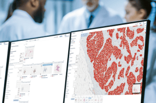
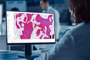
The Suites are a portfolio of tools containing: a clinical viewer, platform for QC and adaptation and an AI model for diagnostic support. Explore our already CE-IVD marked tools and what’s to come!

Blog Image - ER cells with viewer
Scroll down to read more
Only certain Aiforia® Clinical AI models and the Aiforia® Clinical Suite Viewer are CE-IVD marked for diagnostic use in EU and EEA countries, see here for the full list: www.aiforia.com/aiforia-clinical-suite. In all other countries, the use is limited to Research Use Only, not for use in diagnostic procedures.

Getting started is straightforward

Each Clinical Suite can be seamlessly integrated into any existing lab setup: PACS, LIS, scanning, staining, scoring definitions, and more! You don’t need to adapt to start using Aiforia’s solutions, we adapt to meet your needs.

Watch this video to see how Aiforia Clinical AI models are integrated into a clinical pathology lab:

THE AIFORIA CLINICAL SUITES: AI MODELS

Explore our CE-IVD marked clinical AI models:

One of the key tools in the portfolio of our Aiforia Clinical Suites is our repertoire of clinical AI models for diagnostic support.

PR and Gleason
Prostate Cancer: Gleason

Aiforia® Clinical AI Model for Prostate Cancer; Gleason Grade Groups

  • Rapidly distinguishes between normal and cancerous tissue supporting the pathologist in the quick detection of tumor areas
  • Automatically scores a quantitative Gleason grade score and grade group from WSI, saving the pathologist time
  • Provides visual feedback as it allows to view images in different magnifications, move in different x-y-z-locations, view image analysis results, and mark and measure significant features in the images
PR-1
Breast Cancer: PR

Aiforia® Clinical AI Model for Breast Cancer; PR

  • Creates a heatmap assisting the pathologist in automatically finding the critical areas of the sample
  • Rapidly distinguishes between normal and cancerous tissue supporting the pathologist in the quick detection of tumor epithelium
  • Automatically scores the PR positive and negative cells of epithelial origin from either WSI or selected image areas, saving the pathologist time
  • Provides intelligent viewing as the PR AI Model enables viewing and selection of areas with high density of PR-positive cells, or hotspots, displaying the results of image analysis
Clinical_ER_tab
Breast Cancer: ER

Aiforia® Clinical AI Model for Breast Cancer; ER

  • Creates a heatmap assisting the pathologist in automatically finding the critical areas of the sample.

  • Rapidly distinguishes between normal and cancerous tissue supporting the pathologist in the quick detection of tumor epithelium.

  • Automatically scores the ER positive and negative cells of epithelial origin from either WSI or selected image areas, saving the pathologist time.

  • Provides intelligent viewing as the ER AI Model enables viewing and selection of areas with high density of ER-positive cells, or hotspots, displaying the results of image analysis.

Clinical_Ki67_tab
Breast Cancer: Ki67

Aiforia® Clinical AI Model for Breast Cancer; Ki67

  • Creates a heatmap assisting the pathologist in automatically finding the critical areas of the sample.

  • Rapidly distinguishes between normal and cancerous tissue supporting the pathologist in the quick detection of tumor epithelium.

  • Automatically scores the Ki67 positive and negative cells of epithelial origin from either WSI or selected image areas, saving the pathologist time.

  • Provides intelligent viewing as the Ki67 AI Model enables viewing and selection of areas with high density of Ki67-positive cells, or hotspots, displaying the results of image analysis.

Clinical_PDL1_tab
Lung Cancer: PD-L1

Aiforia® Clinical AI Model for Lung Cancer; PD-L1

  • Creates a heatmap assisting the pathologist in automatically finding the critical areas of the sample.

  • Rapidly distinguishes between normal and cancerous tissue supporting the pathologist in the quick detection of tumor epithelium.

  • Automatically scores the PD-L1 positive and negative cells of epithelial origin from whole slide images (WSI) saving the pathologist time when evaluating patient samples.

  • Provides intelligent viewing and selection of areas with high density of PD-L1 positive cells displaying the results of image analysis in different magnifications and enables the user to move in different x-y-z-locations, as well as to mark and measure features in the images.

A MULTITUDE OF CLINICAL POSSIBILITIES IN A SINGLE, UNIFIED PLATFORM

Each Aiforia Clinical Suite offers the tools you need to transform your clinical workflow:

Aiforia Clinical AI Model for Diagnostic Support

  • The deep learning AI model provides fast and powerful image analysis assistance to pathologists in their diagnostic tasks
  • CE-IVD marked Aiforia Clinical AI Models for Breast Cancer: Ki67, ER, and PR available now
  • CE-IVD marked Aiforia Clinical AI Model for Lung Cancer: PD-L1 available now
  • CE-IVD marked Aiforia Clinical AI Model for Prostate Cancer: Gleason Grade Groups available now
  • Improves diagnostic accuracy, reduces bias and standardizes analysis in sample review
  • Speeds up time-consuming tasks to free up more time for rare and complex cases
  • Increasing diagnostic confidence
  • The Aiforia clinical AI model can also be used for pre- and post-analysis screening, see below for these additional features

CE-IVD marked Aiforia® Clinical Suite Viewer

  • Rapidly displays AI-assisted analysis results in an intuitive and easily readable format and enabling case prioritization based on severity
  • Automatically generates reports from analyses by clinical AI models, significantly saving the pathologist time and improving the lab’s overall output efficiency
  • Supports full interaction with the pathologist by allowing viewing of images in different magnifications, movement in different x-y-z-locations, viewing of image analysis results in high precision, and markings and measurement of significant features in the images
  • Users can easily edit the results in the report, select hotspot areas and exclude control tissue or areas of bad quality

Find out more about the Viewer.

Aiforia Platform for QC and adaptation

  • An intuitive user interface in which you can easily adapt your AI model to accommodate changes if they are needed
  • Easy management and monitoring for QC and auditing purposes
  • Compliant with the FDA’s Good Machine Learning Practices (GMLP), a framework outlining best practices in software engineering and quality management of AI systems
Additionally

You can use the Aiforia Clinical AI model for:

PRE-ANALYSIS SCREENING

Improve lab and workflow efficiency

  • Automatically prioritize cases for the pathologist to focus on the most severe ones first
  • Speed up time to diagnosis and detect more cases 

POST-ANALYSIS SCREENING

Improve patient safety

  • To act as a safety net by confirming the decisions of the pathologist and alerting them to potentially suspicious findings
  • Enabling the detection of more cases and reducing potential errors

Are you ready to experience the power of AI?

We are looking for pathology labs to join us in becoming the frontrunners of AI-powered diagnostics.

Benefits

Work more efficiently

Diagnose more patients in less time and speed up case review

ic_slideincrease_white

Improve accuracy

Improve diagnostic accuracy and be confident in your decision making

ic_define_white

Reduce bias

and standardize sample review therefore democratizing care

ic_reducebias_white

Harness your full potential

Unbind yourself from time-consuming, manual and frustrating tools and processes

Free your time

Allow yourself more time to focus on rare and complex cases

ic_save time_white

Increase output

Detect more cases in less time, take on more patients

ic_output_white-1

Improve patient outcomes

Precise diagnosis permits efficient and personalized therapies to be administered

ic_patientoutcome_white

Enhance service quality

Shorten time to diagnosis and reduce missed diagnoses and misdiagnoses

ic_decisions with confidence_white

Save on cost and time

Significant time savings, 90% less hands-on time for pathologists and reduce misdiagnosis and error while improving treatment accuracy

ic_reduce costs_white

Enhance staff satisfaction

Better workload distribution can be achieved as pathologists spend less time on manual, repetitive tasks

ic_smile_white

Less time waiting

Get your diagnosis and results faster

ic_save time_white

Effective treatment

With a precise diagnosis clinicians are able to treat more effectively

ic_personalcare_white

Democratized care

Samples are reviewed consistently and efficiently ensuring everyone is treated the same

ic_collaborate remotly_white

Reduction in unnecessary interventions

Reducing missed and misdiagnosis means a reduction in the likelihood of unnecessary surgeries or procedures

ic_regression_white

ON-DEMAND WEBINAR

Aiforia’s CE-IVD marked clinical AI models for Ki67 and PD-L1

Watch this recording for an intro and demo from pathologists Dr. Laury and Dr. Sjöblom on the Aiforia® Clinical AI Model for Lung Cancer; PD-L1 and Aiforia® Clinical AI Model for Breast Cancer; Ki67. 

Watch now
aiforia_bg_1
Seeing is believing
Book a free demonstration!
Book a demo