Breast Cancer Suite

Automates breast cancer grading from H&E-stained whole-slide images (WSI), accurately identifying invasive carcinoma and ductal carcinoma in situ (DCIS). It objectively scores mitotic count, tubular formation, and nuclear pleomorphism, addressing key challenges of manual grading such as variability and time constraints, consistent with the Nottingham Grading System.
Clinical performance highlights
- Substantially reduces grading time (50% faster)
- High correlation with expert grading assessments
- Consistently reproducible, reducing interobserver variability
CE-IVD marked for diagnostic use in EU and EEA countries (IVDR) and for Research Use Only (RUO) and Performance Studies Only (PSO) in all other market areas

Automatically detects invasive carcinoma and quantifies ER-positive and negative tumor cells from whole-slide images (WSI) or selected tissue areas, supporting consistent and objective scoring.
Clinical performance highlights
- Exceptional diagnostic accuracy with high correlation to expert assessments
- Improved diagnostic consistency with reduced interobserver variability
- Accelerated workflow with up to 47% faster ER scoring
CE-IVD marked for diagnostic use in EU and EEA countries, and for Research Use Only (RUO) and Performance Studies Only (PSO) in all other market areas

Automatically detects invasive carcinoma and quantifies PR-positive and negative cells from whole-slide images (WSI) or selected tissue regions, enhancing diagnostic clarity and workflow.
Clinical performance highlights
- High diagnostic confidence with enhanced interobserver agreement
- Reliable reproducibility with near-perfect consistency
- Efficient workflow with approximately 12% faster scoring
CE-IVD marked for diagnostic use in EU and EEA countries, and for Research Use Only (RUO) and Performance Studies Only (PSO) in all other market areas

Accurately identifies invasive carcinoma and scores HER2 expression in alignment with CAP guidelines, improving diagnostic reliability and workflow efficiency.
Clinical performance highlights
- Outstanding sensitivity and precision for confident HER2 classification
- Enhanced diagnostic consistency with increased interobserver concordance
- Objective support to identify challenging HER2-low and HER2-ultralow cases
CE-IVD marked for diagnostic use in EU and EEA countries (IVDR) and for Research Use Only (RUO) and Performance Studies Only (PSO) in all other market areas

Automatically detects invasive carcinoma and quantifies Ki67-positive and negative tumor cells from whole-slide images (WSI) or targeted image regions, improving diagnostic accuracy and workflow.
Clinical performance highlights
- High diagnostic accuracy with excellent correlation to expert visual scoring
- Enhanced diagnostic concordance through improved pathologist agreement
- Significant time savings, reducing scoring duration by up to 54%
- Reliable precision with automated hotspot detection for accurate proliferation assessment
CE-IVD marked for diagnostic use in EU and EEA countries, and for Research Use Only (RUO) and Performance Studies Only (PSO) in all other market areas

Supports the detection and quantification of metastases of breast cancer in lymph nodes from whole slide images.
Clinical performance highlights
- Showed excellent correlation with visual diagnosis
- Saved time (up to 40% faster)
Aiforia® Lymph Node Metastasis is currently for Research Use Only (RUO) and for Performance Studies Only (PSO) in all market areas.
Breast Cancer Suite
Aiforia® Breast Cancer Grading

Automates breast cancer grading from H&E-stained whole-slide images (WSI), accurately identifying invasive carcinoma and ductal carcinoma in situ (DCIS). It objectively scores mitotic count, tubular formation, and nuclear pleomorphism, addressing key challenges of manual grading such as variability and time constraints, consistent with the Nottingham Grading System.
Clinical performance highlights
- Substantially reduces grading time (50% faster)
- High correlation with expert grading assessments
- Consistently reproducible, reducing interobserver variability
CE-IVD marked for diagnostic use in EU and EEA countries (IVDR) and for Research Use Only (RUO) and Performance Studies Only (PSO) in all other market areas
Aiforia® Breast Cancer ER

Automatically detects invasive carcinoma and quantifies ER-positive and negative tumor cells from whole-slide images (WSI) or selected tissue areas, supporting consistent and objective scoring.
Clinical performance highlights
- Exceptional diagnostic accuracy with high correlation to expert assessments
- Improved diagnostic consistency with reduced interobserver variability
- Accelerated workflow with up to 47% faster ER scoring
CE-IVD marked for diagnostic use in EU and EEA countries, and for Research Use Only (RUO) and Performance Studies Only (PSO) in all other market areas
Aiforia® Breast Cancer PR

Automatically detects invasive carcinoma and quantifies PR-positive and negative cells from whole-slide images (WSI) or selected tissue regions, enhancing diagnostic clarity and workflow.
Clinical performance highlights
- High diagnostic confidence with enhanced interobserver agreement
- Reliable reproducibility with near-perfect consistency
- Efficient workflow with approximately 12% faster scoring
CE-IVD marked for diagnostic use in EU and EEA countries, and for Research Use Only (RUO) and Performance Studies Only (PSO) in all other market areas
Aiforia® Breast Cancer HER2

Accurately identifies invasive carcinoma and scores HER2 expression in alignment with CAP guidelines, improving diagnostic reliability and workflow efficiency.
Clinical performance highlights
- Outstanding sensitivity and precision for confident HER2 classification
- Enhanced diagnostic consistency with increased interobserver concordance
- Objective support to identify challenging HER2-low and HER2-ultralow cases
CE-IVD marked for diagnostic use in EU and EEA countries (IVDR) and for Research Use Only (RUO) and Performance Studies Only (PSO) in all other market areas
Aiforia® Breast Cancer Ki67

Automatically detects invasive carcinoma and quantifies Ki67-positive and negative tumor cells from whole-slide images (WSI) or targeted image regions, improving diagnostic accuracy and workflow.
Clinical performance highlights
- High diagnostic accuracy with excellent correlation to expert visual scoring
- Enhanced diagnostic concordance through improved pathologist agreement
- Significant time savings, reducing scoring duration by up to 54%
- Reliable precision with automated hotspot detection for accurate proliferation assessment
CE-IVD marked for diagnostic use in EU and EEA countries, and for Research Use Only (RUO) and Performance Studies Only (PSO) in all other market areas
Aiforia® Lymph Node Metastasis

Supports the detection and quantification of metastases of breast cancer in lymph nodes from whole slide images.
Clinical performance highlights
- Showed excellent correlation with visual diagnosis
- Saved time (up to 40% faster)
Aiforia® Lymph Node Metastasis is currently for Research Use Only (RUO) and for Performance Studies Only (PSO) in all market areas.
AI for breast cancer diagnostics
Aiforia® Breast Cancer Suite offers a fully digital cockpit for breast cancer diagnostics, supporting the entire diagnostic workflow.
Lung Cancer Suite

Automatically detects invasive carcinoma and delivers standardized, reproducible TPS scoring, enhancing diagnostic accuracy and efficiency. It assists pathologists in overcoming the challenges of subjective and time-consuming manual PD-L1 scoring in lung cancer.
Clinical performance highlights
- Excellent diagnostic accuracy with high correlation to expert visual assessments
- Enhanced consistency through improved pathologist concordance (Fleiss’ Kappa ~0.87)
- Significantly streamlined workflow, reducing analysis time by up to 46%
- Reliable and standardized scoring at critical clinical thresholds (e.g., 1% and 50%)
CE-IVD marked for diagnostic use in EU and EEA countries, and for Research Use Only (RUO) and Performance Studies Only (PSO) in all other market areas
Lung Cancer Suite
Aiforia® Lung Cancer PD-L1

Automatically detects invasive carcinoma and delivers standardized, reproducible TPS scoring, enhancing diagnostic accuracy and efficiency. It assists pathologists in overcoming the challenges of subjective and time-consuming manual PD-L1 scoring in lung cancer.
Clinical performance highlights
- Excellent diagnostic accuracy with high correlation to expert visual assessments
- Enhanced consistency through improved pathologist concordance (Fleiss’ Kappa ~0.87)
- Significantly streamlined workflow, reducing analysis time by up to 46%
- Reliable and standardized scoring at critical clinical thresholds (e.g., 1% and 50%)
CE-IVD marked for diagnostic use in EU and EEA countries, and for Research Use Only (RUO) and Performance Studies Only (PSO) in all other market areas
Prostate Cancer Suite

Automatically identifies adenocarcinoma, streamlines the Gleason grading process, and directly addresses interobserver variability and subtle pattern detection challenges commonly faced by pathologists.
Clinical performance highlights
- Exceptional diagnostic accuracy with excellent correlation to expert grading
- Improved reproducibility and reduced interobserver variability
- Significantly faster Gleason scoring, saving up to 34% of analysis time
CE-IVD marked for diagnostic use in EU and EEA countries, and for Research Use Only (RUO) and Performance Studies Only (PSO) in all other market areas

Efficiently detects subtle perineural invasion (PNI) features, addressing the common diagnostic challenge of accurately and consistently identifying tumor involvement around nerves.
Clinical performance highlights
- High diagnostic confidence with excellent correlation to visual assessments
- Improved reproducibility, ensuring reliable identification of subtle PNI
- Enhanced workflow efficiency with scoring times reduced by up to 29%
CE-IVD marked for diagnostic use in EU and EEA countries (IVDR) and for Research Use Only (RUO) and Performance Studies Only (PSO) in all other market areas

Automatically detects Gleason Grade 4 Cribriform patterns in prostate biopsy whole slide images (WSI), addressing pathologists' challenges with subjective, time-consuming visual assessments, with the aim to reduce diagnostic variability and enhance workflow efficiency.
Clinical performance highlights
- Excellent diagnostic reliability, consistently aligning with expert visual assessments
- Improved reproducibility and reduced diagnostic variability
- Significantly reduced analysis time, improving workflow by up to 54%
CE-IVD marked for diagnostic use in EU and EEA countries (IVDR) and for Research Use Only (RUO) and Performance Studies Only (PSO) in all other market areas
Prostate Cancer Suite
Aiforia® Prostate Cancer Gleason Grade Groups

Automatically identifies adenocarcinoma, streamlines the Gleason grading process, and directly addresses interobserver variability and subtle pattern detection challenges commonly faced by pathologists.
Clinical performance highlights
- Exceptional diagnostic accuracy with excellent correlation to expert grading
- Improved reproducibility and reduced interobserver variability
- Significantly faster Gleason scoring, saving up to 34% of analysis time
CE-IVD marked for diagnostic use in EU and EEA countries, and for Research Use Only (RUO) and Performance Studies Only (PSO) in all other market areas
Aiforia® Prostate Cancer PNI

Efficiently detects subtle perineural invasion (PNI) features, addressing the common diagnostic challenge of accurately and consistently identifying tumor involvement around nerves.
Clinical performance highlights
- High diagnostic confidence with excellent correlation to visual assessments
- Improved reproducibility, ensuring reliable identification of subtle PNI
- Enhanced workflow efficiency with scoring times reduced by up to 29%
CE-IVD marked for diagnostic use in EU and EEA countries (IVDR) and for Research Use Only (RUO) and Performance Studies Only (PSO) in all other market areas
Aiforia® Prostate Cancer G4 Cribriform

Automatically detects Gleason Grade 4 Cribriform patterns in prostate biopsy whole slide images (WSI), addressing pathologists' challenges with subjective, time-consuming visual assessments, with the aim to reduce diagnostic variability and enhance workflow efficiency.
Clinical performance highlights
- Excellent diagnostic reliability, consistently aligning with expert visual assessments
- Improved reproducibility and reduced diagnostic variability
- Significantly reduced analysis time, improving workflow by up to 54%
CE-IVD marked for diagnostic use in EU and EEA countries (IVDR) and for Research Use Only (RUO) and Performance Studies Only (PSO) in all other market areas
Aiforia® Prostate Cancer HG-PIN

Supports the detection of high-grade prostatic intraepithelial neoplasia (high-grade PIN) in whole slide images (WSI) of prostate tissue.
Aiforia® Prostate Cancer HG-PIN is currently for Research Use Only (RUO) and for Performance Studies Only (PSO) in all market areas.
Colon Suite

Identifies important histological features of colorectal cancer and provides a recurrence prediction estimate useful for treatment decisions. This prognostic AI model was developed and validated in collaboration with the Mayo Clinic.
Clinical performance highlights
- Verified and validated in the Aiforia® Platform using whole slide images from eight independent hospitals and patient cohorts
- Improved risk stratification as compared to the American Society of Clinical Oncology staging
Aiforia® Colorectal Cancer QuantCRC is currently for Research Use Only (RUO) and for Performance Studies Only (PSO) in all market areas.

Supports the detection and quantification of metastases of colorectal cancer in lymph nodes from whole slide images. The AI model was developed in collaboration with the University of Bern.
Clinical performance highlights
- Showed excellent correlation with visual diagnosis
- Saved time (up to 40% faster)
Aiforia® Lymph Node Metastasis is currently for Research Use Only (RUO) and for Performance Studies Only (PSO) in all market areas.
Colon Suite
Aiforia® Colorectal Cancer QuantCRC

Identifies important histological features of colorectal cancer and provides a recurrence prediction estimate useful for treatment decisions. This prognostic AI model was developed and validated in collaboration with the Mayo Clinic.
Clinical performance highlights
- Verified and validated in the Aiforia® Platform using whole slide images from eight independent hospitals and patient cohorts
- Improved risk stratification as compared to the American Society of Clinical Oncology staging
Aiforia® Colorectal Cancer QuantCRC is currently for Research Use Only (RUO) and for Performance Studies Only (PSO) in all market areas.
Aiforia® Lymph Node Metastasis

Supports the detection and quantification of metastases of colorectal cancer in lymph nodes from whole slide images. The AI model was developed in collaboration with the University of Bern.
Clinical performance highlights
- Showed excellent correlation with visual diagnosis
- Saved time (up to 40% faster)
Aiforia® Lymph Node Metastasis is currently for Research Use Only (RUO) and for Performance Studies Only (PSO) in all market areas.
Gastric Suite
Gastric Suite
Aiforia® Gastric Cancer

Supports the detection of gastric cancer and Helicobacter pylori in whole slide images (WSI) of H&E-stained FFPE gastric tissue.
Aiforia® Gastric Cancer is currently for Research Use Only (RUO) and for Performance Studies Only (PSO) in all market areas.
Lymph Node Metastasis

Supports the detection and quantification of metastases of breast cancer, melanoma, and colorectal cancer in lymph nodes from whole slide images.
Clinical performance highlights:
- Showed excellent correlation with visual diagnosis
- Saved time (up to 40% faster)
Aiforia® Lymph Node Metastasis is currently for Research Use Only (RUO) and for Performance Studies Only (PSO) in all market areas.
Lymph Node Metastasis
Aiforia® Lymph Node Metastasis

Supports the detection and quantification of metastases of breast cancer, melanoma, and colorectal cancer in lymph nodes from whole slide images.
Clinical performance highlights:
- Showed excellent correlation with visual diagnosis
- Saved time (up to 40% faster)
Aiforia® Lymph Node Metastasis is currently for Research Use Only (RUO) and for Performance Studies Only (PSO) in all market areas.
Elevate diagnostic capabilities with Aiforia® Clinical Suites

Complete AI-driven workflow
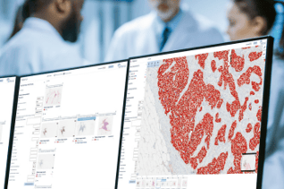
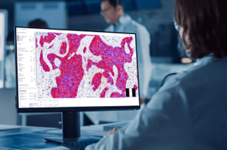
Aiforia’s solutions support the complete AI-driven workflow through an optimized combination of the worklist, interactive Clinical Viewer, a broad portfolio of quantitative AI models, and case-level reporting.

Comprehensive case-level review and reporting
Aiforia® Clinical Suites conveniently combine results from multiple AI models, WSIs, and stainings into a case-level view and report for first-read diagnostic support.

Precise, transparent AI results
Aiforia takes accuracy beyond standard AI heatmaps by clearly marking the exact findings with detailed visual overlays directly on the digital slides, enabling easy visual confirmation at any magnification level. This approach empowers pathologists with both visual transparency and exact quantitative data, delivering actionable insights.

Seamless integrations & flexible deployment
Aiforia offers seamless integration into existing laboratory systems, with publicly available API specifications. Customers can choose from different deployment options according to their needs.

The benefits of AI in clinical diagnostics
Increased efficiency
Diagnose more patients in less time by unbinding yourself from time-consuming, manual tasks.
Advanced precision
Improve diagnostic accuracy and be confident in your decision-making.
Improved consistency
Reduce variability to standardize your sample review and ensure democratized patient care.
AI for clinical diagnostics
WEBINAR
PD-L1 on the edge: cut-offs, cognitive bias & the case for AI in lung cancer pathology
This 45-minute webinar takes you inside the real-world diagnostic pressure points of PD-L1 TPS testing in NSCLC. With insights drawn from clinical research, pathology practice, and oncology decision-making, we examine the human limits of PD-L1 scoring and how AI can sharpen clinical decisions.
BLOG POST
How did the Mayo Clinic choose a vendor for AI in pathology?
What to consider when choosing a vendor for AI in pathology? Learn how the Mayo Clinic evaluated platform providers based on six key criteria.
BLOG POST
Digital pathology deployment in Veneto: 8 steps to success
What is required for pathology laboratories to go fully digital? Learn how the broadest public healthcare provider in the Veneto region did it.
BLOG POST
5 reasons to use AI in clinical diagnostics
What additional benefit does AI bring to the digitized pathology workflow in clinical diagnostics? Read our article to find out.
INTERVIEW
Benefits of AI and digital pathology to the clinical lab
Clinical pathologists, Dr. Kevin Sandeman and Dr. Tuomas Mirtti, discuss the impact and benefits of AI and digital pathology in clinical diagnostics.
Request a demo
Discover the power of AI for image analysis
Find out how to enhance your image analysis work in diagnostic pathology, preclinical studies, or medical research. The demo will be tailored based on your interests.
The demo will help you understand:
- How AI-assisted image analysis can increase efficiency, precision, and consistency in the pathology workflow.
- The limitless possibilities Aiforia® Platform offers – both for research and clinical diagnostics – and the suitable use cases for your needs.
We can either demonstrate Aiforia’s image analysis solutions on your own images or any of our 400+ application examples (i.e., neuron quantification, automated tumor grading, NASH analysis, etc.).
Fill in the form, and one of our experts will contact you shortly to schedule the time.