
Clinical

  • Clinical Solutions
  • AI solutions portfolio

Explore Aiforia’s comprehensive portfolio of high-performance AI solutions, engineered for both clinical and preclinical applications. Aiforia’s solutions aim to enhance the speed, accuracy, and consistency of analyzing large and complex medical images, especially in pathology.

Learn more

Research

  • Research solutions
  • Aiforia® Create
  • AI solutions portfolio

Aiforia’s research solutions offer fully automated study-centric workflows and pathology image analysis applications that cater to all research areas. Empowered by robust AI capabilities, pathologists and scientists can make discoveries while saving time by automating repetitive tasks – also within the framework of GLP. Aiforia’s solutions can be integrated with any existing laboratory infrastructure to enjoy the full benefits of a digitized workflow.

Learn more

Aiforia® Create is the most versatile tool for developing deep learning AI models for image analysis in digital pathology. Its cloud-based, collaborative working environment allows multiple users to work together in real time, anywhere in the world. Praised for its intuitive user interface, it allows users a fast start, even without any prior AI experience.Learn more >

Learn more

Explore Aiforia’s comprehensive portfolio of high-performance AI solutions, engineered for both clinical and preclinical applications. Aiforia’s solutions aim to enhance the speed, accuracy, and consistency of analyzing large and complex medical images, especially in pathology.

Learn more

AI development tool

  • Aiforia® Create

Aiforia® Create is the most versatile tool for developing deep learning AI models for image analysis in digital pathology. Its cloud-based, collaborative working environment allows multiple users to work together in real time, anywhere in the world. Praised for its intuitive user interface, it allows users a fast start, even without any prior AI experience.

Learn more

Resources

  • Publications
  • Resource Library
  • Press releases

Explore some of our latest publications showcasing the discoveries enabled by the Aiforia® Platform.

Learn more

Aiforia's insights and resources for AI in pathology. View some of our content pieces from the world of AI.

Learn more

Stay updated with Aiforia’s recent announcements. Explore our latest news, updates and announcements.

Learn more

Company

  • About us
  • Investors
  • Partners
  • Media
  • Events
  • Careers
  • Quality and security

Find out more about shareholder matters, investment discussions, and financial-related information.

Learn more

Aiforia's mission is to transform pathology image analysis with AI, enabling better care for each patient. To succeed, we need our trusted partners. Explore Aiforia’s partner network.

Learn more

Experience our AI solutions firsthand. Explore the list of conferences where you can connect with Aiforia’s team live and book a time to discover the future of AI-powered image analysis.

Learn more

Our mission is to transform pathology image analysis with AI, enabling better care for each patient. Find out more about job openings, thesis opportunities, or submitting applications.

Learn more

Aiforia Technologies holds information security to the highest standard. We hold several, globally relevant quality and security certifications, so you can be sure your sensitive information is safe.

Learn more

Research

Research solutions

Aiforia® Create

AI solutions portfolio

AI development tool

Aiforia® Create

Resources

Publications

Resource Library

Press releases

Van Andel Institute case study: quantitative assessment of alpha-synuclein immunoreactivity with AI

Van Andel Institute created four AI models with Aiforia to provide unbiased, quantitative analysis for Parkinson’s disease research.
Written by Aiforia

We interviewed Christopher Tulisiak, PhD, a Postdoctoral Fellow at the Van Andel Institute, about their research on Parkinson's disease. Read his answers below. 

Why did you decide to use Aiforia’s AI software and services? 

"We recognized that Aiforia could provide us a uniform, fully quantitative, unbiased way to analyze the presence of a specific marker of pathogenic α-synuclein, a key hallmark of Parkinson’s disease. We had previously used several different analysis methods to assess pathogenic α-synuclein immunoreactivity in the brain, ranging from semi-quantitative scoring to densitometry to stereology.

None were perfect, either lacking complete objectivity, requiring perfect staining and imaging, or quantifying only Lewy bodies, but not the highly abundant and stringy Lewy neurites. Because Lewy neurites are highly abundant in our mouse model, but as no analysis method was able to perfectly represent them quantitatively, a large portion of pathology was missed by these tools."

For which projects was Aiforia used?

"Our project has utilized Aiforia’s custom AI services, which have seen us work closely with Aiforia's outstanding Field Application Specialist (FAS) to develop four separate AI models that contribute either to quantitating the number or morphology of specific cell types or to the presence of pathogenic α-synuclein."  
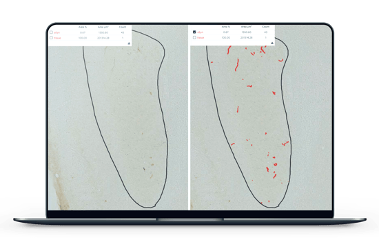
alpha-synuclein immunoreactivity with AI

"Our FAS has worked diligently to train the AI models based on our criteria, and then members of our laboratory work as external validators of each of the AI models’ performance. After each validation, the AI is fine-tuned into a new iteration until it is ready for release when it performs with an exceptionally low error rate in comparison to validators’ annotations."
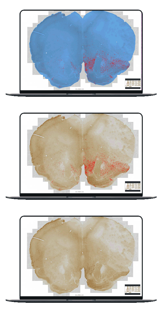
alpha-synuclein immunoreactivity with AI

"We will soon be transitioning to Aiforia® Create in order to continue to fine-tune our AI models when we have sections that vary from those on which the AI was trained."

What were the other three Aiforia AI models trained to detect and analyze?

"Using the different tools Aiforia provides, we were able to develop AI models for the quantitation of both tyrosine hydroxylase (TH)-positive neurons (which are selectively lost in Parkinson’s disease) by cell counts, and astrocyte and microglia activation (which is a common immunoreaction to pathological insults) by morphological analysis. 

Aiforia's software saves us many hours of work because, beyond imaging (which is now passively achieved using a scanner, as opposed to actively with a microscope), analysis is automated by the AI model over the course of minutes (or hours at most). After the analysis is complete, we export the results, organize them using our statistics software of choice, and generate figures."

What is the biggest benefit to your research?

"Our AI models provide the objectivity of fully quantitative analysis that covers entire brain regions and time-saving through passive image acquisition and analysis, during which we can accomplish other goals.

For the TH model, coverage is greatly increased and practically more accurate than an unbiased sampling of stereology (which is excessively time-consuming). For astrocyte and microglia models, the goal is higher accuracy compared to contrast-based densitometry (and increased coverage). And for α-synuclein, we capture a full representation of varying synucleinopathy morphologies in a fully quantitative manner."

Read more neuroscience case studies: