Ashley Scherlek, a research technician at Massachusetts General Hospital (MGH) working on Alzheimer’s disease, delegates her image analysis tasks to an artificial intelligence model she created. Ashley is not a data scientist, nor does she have previous experience with AI. She used Aiforia’s AI development software, Aiforia® Create, to train neural networks to learn how to identify and quantify cells and cellular deposits in brain tissue samples at the Harvard Medical School hospital where she is working.
To utilize supervised artificial intelligence for image analysis or recognition, the neural networks that form AI must be trained to recognize and detect the feature or area of interest. With Aiforia Create, this training simply requires the user to annotate their images, thereby teaching the neural networks what to look for and ultimately producing an AI model the user can then use to automate that task.
Getting started with Aiforia Create
The first step in getting started was for Ashley to upload her images to the Aiforia® Platform, which supports any 2D image, including brightfield and fluorescence (IF). Ashley made sure to choose images that represented variability across the entire dataset.
While Aiforia’s tools are built on an intuitive user interface, all users receive training and support throughout the whole process to ensure ease and efficiency in AI model development. Aiforia’s in-house scientific experts run a six to eight-week training plan with each user, offering them a mixture of real-time training and supportive materials, resources, and guides. The training encompasses the entire AI model development process.
“During my virtual lessons, Aiforia’s science team did an exceptional job going through all the details of my projects and giving me the tools to train the neural networks on my own,” Ashley explains. She was supported by Aiforia’s Field Application Scientist, who has a strong background in studying Alzheimer’s disease.
Training the neural networks
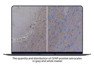
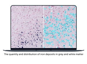 Images were uploaded and securely stored, the bulk of the training was complete, and Ashley was able to quickly get started on working with the neural networks in Aiforia Create. To train them she used approximately 20 slides per training dataset and annotated about 3% of the tissue area. Ashley’s goal was to train the neural networks to detect and quantify reactive astrocytes and iron deposits, ultimately producing two separate AI models.
“I’ve loved using Aiforia Create. The whole team has been very responsive and given me the support to freely create my projects,” Ashley explained after her onboarding and AI model training was complete. Despite the complexity of her project, which consisted of multiple feature layers containing unique classes, Ashley was able to train and produce within just a few weeks two robust AI models to assist her and the MGH lab in assessing a neurological condition called cortical superficial siderosis (cSS), as a neuropathological correlate of vascular disease.
Deploying the AI models
 Ashley’s two AI models are now rapidly and unbiasedly performing her manual, time-consuming tasks for her, as she explains:
“Using AI for image analysis allows us to generate more quantitative data and to perform more advanced statistical analysis compared to previous semi-quantitative image analysis approaches that I used in the past.”
Ashley’s project and experience are not unique but normal for all Aiforia Create users for applications in a huge variety of fields. The AI development software supports all image files, formats, and scanner types. The neural networks can then be trained to detect, quantify, measure, and analyze anything. If you can see it, you can train the AI to find it.
Read more neuroscience case studies: