

 Back to all blog posts


Ki-67 is an important breast cancer (BC) marker, especially for adjuvant treatment in HR+, HER2- cases. Working groups have provided guidance for Ki-67 immunohistochemistry (IHC) BC scoring to limit pathologist’s variability, but no scoring method has been universally accepted. Rapid and reliable image analysis solutions to support scoring have surfaced for the Ki-67 assessment.
In this study, a team from Cerba Research compared the results of Ki-67 scoring performed with the Aiforia® Platform (deep learning Al platform) against two independent pathologists in a breast cancer population in terms of scoring quality and time savings.
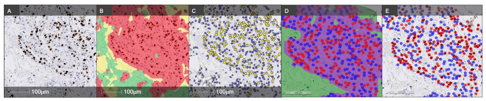
Figure 1. Image analysis illustration. From left to right: Ki-67 IHC, DAB detection (brown), hematoxylin counterstain (A). The Halo classifier with the tumor area in red, the non-tumor area in green and the background in yellow (B). Halo analysis markup Ki-67, blue: nuclei and yellow: positive cells (C). Aiforia tissue detection with the tumor area in purple and the nontumor area in green (D). Aiforia analysis markup Ki-67, blue: negative cells and red: positive cells (E). Scale bar 100μm.
The workflow
The team stained 114 breast cancer tumors for Ki-67 (Ki-67 clone MIB-1, ref GA626-Agilent) on the Dako Omnis platform. Three methodologies were used to quantify Ki-67+ tumor cells:
- Aiforia’s deep learning model, Aiforia® Breast Cancer Ki67, was calibrated to work on the images of this case.
- Two pathologists (Patho 1 and Patho 2) were trained following the International Ki67 Working Group (IKWG) guidelines1,2. Intra-analysis assessment was done for one pathologist. The selected pathologist re-read the samples after a three-week washout period.
- The random forest classifier from Halo® was used to separate the image into tumor, non-tumor, and background with the pathologist’s approval. After cell segmentation, Ki-67 positivity was assessed by thresholding3.
The time needed to complete the analyses was recorded for each method.
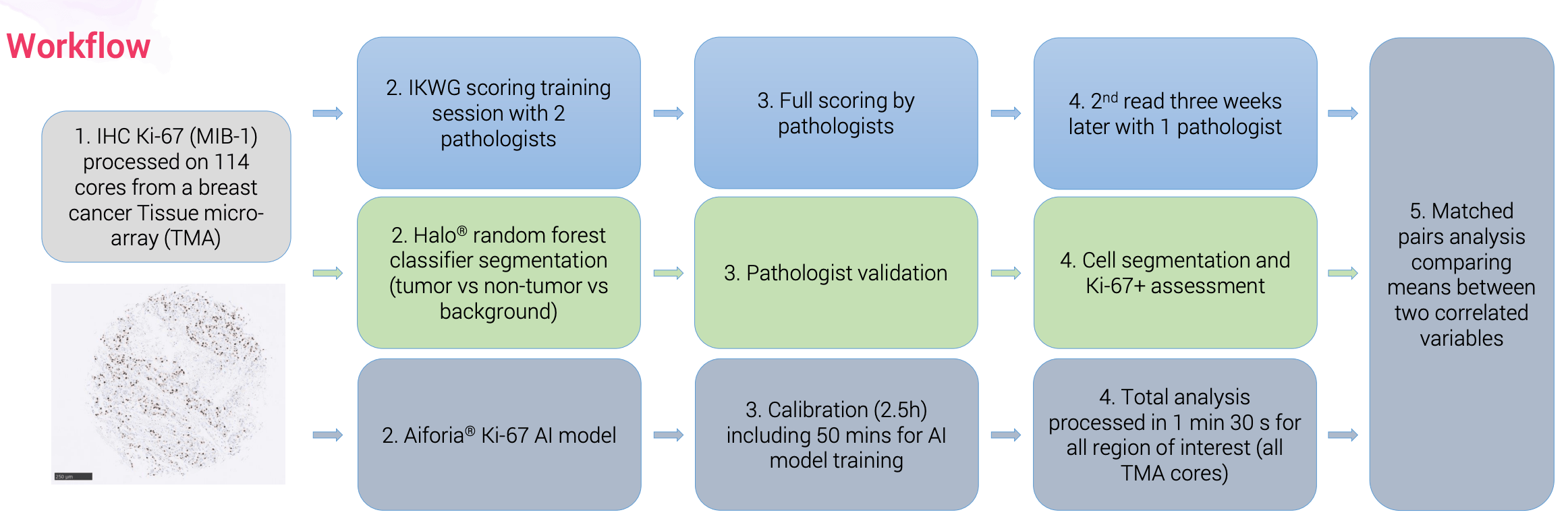
Figure 2. Example of an IHC Ki-67 staining workflow from a breast cancer specimen (invasive carcinoma)
Key takeaways
Out of 114 cores, only 109 were analyzed due to the absence of tissue and/or pathologists unable to score. Ki-67+ cells were detected in an average of 7.79 – 12.33% of tumor cells, depending on the analysis approach. The study shows a very high consistency of results obtained for Ki-67 scoring between the two image analysis software, Aiforia® and Halo®, on breast tumors analyzed. The correlation obtained between the pathologists was, however, weaker (mean r2=0.86), despite appropriate training and following of guidelines, but remains within an acceptable range.
Table 1. Summary of matched pairs analysis of Ki-67 quantification on breast cancer tumors (n=109). Cell color coding for r2: green >0.90; orange: 0.90 - 0.80; yellow: 0.80 - 0.75
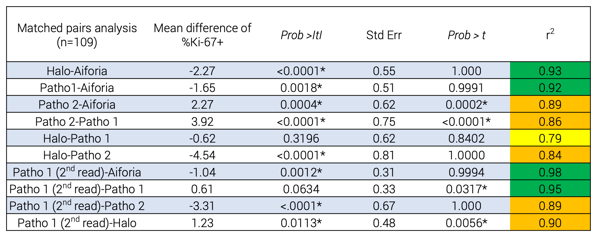
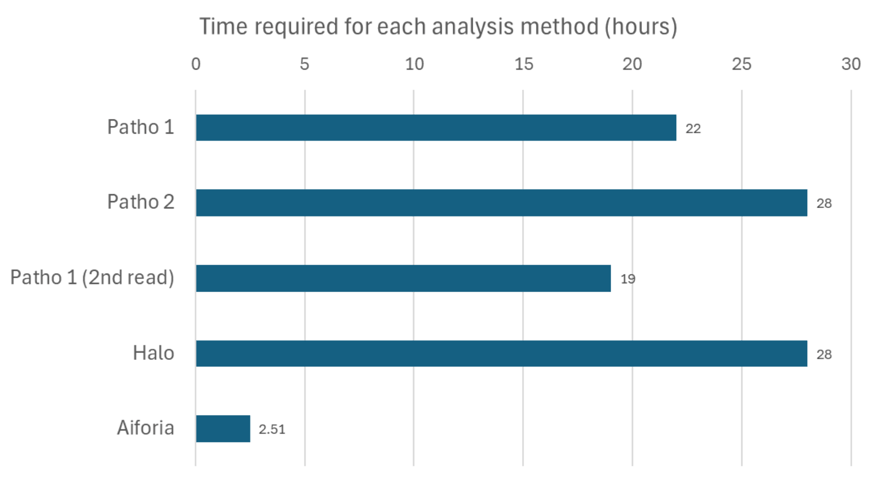
Figure 3. Comparison of the process times required for each method in hours
Aiforia’s deep learning AI approach was by far the quickest, even when including the model training (total time: 2h 51min). Pathologists’ time ranged from 22 to 28 hours without a major gain in analysis time in the second review. Halo® took 28 hours, including application development, pathologist verification, and analysis.
"In a nutshell, the Ki-67 tumor analysis approaches were quite comparable, similar to our previous analysis with the Ki-67 30-9 clone4. These results also demonstrate a significant time benefit of using an AI-driven method for Ki-67 analysis in breast cancer, ensuring that Ki-67 services are delivered efficiently and effectively to patients" – Rania Gaspo, Dir. Global Therapy Area Lead from Cerba Research
The study was presented at ESMO 2024. See the poster here: 175P Utility of artificial intelligence (AI) in Ki67 scoring of a breast cancer (BC) patient population

References
- Polley, M. et al. (2015, June). An international study to increase concordance in Ki-67 scoring. Mod Pathol. 28(6), 778-86. https://doi.org/10.1038/modpathol.2015.38
- Nielsen, T. et al. (2021, July 1). Assessment of Ki-67 in Breast Cancer: Updated Recommendations From the International Ki-67 in Breast Cancer Working Group. J Natl Cancer Inst. 113(7), 808-819. https://doi.org/10.1093/jnci/djaa201
- Welcome to Ki-67-QC calibrator. http://www.gpec.ubc.ca:8080/tmadb-0.1/calibrator/index
- Pichon, X. et al. ESMO 2023. 2329P-Evidence for the utility of artificial intelligence (AI) and image analysis in Ki-67 quantification in solid tumors