
Clinical
Explore Aiforia’s comprehensive portfolio of high-performance AI solutions, engineered for both clinical and preclinical applications. Aiforia’s solutions aim to enhance the speed, accuracy, and consistency of analyzing large and complex medical images, especially in pathology.
Learn moreResearch
Aiforia’s research solutions offer fully automated study-centric workflows and pathology image analysis applications that cater to all research areas. Empowered by robust AI capabilities, pathologists and scientists can make discoveries while saving time by automating repetitive tasks – also within the framework of GLP. Aiforia’s solutions can be integrated with any existing laboratory infrastructure to enjoy the full benefits of a digitized workflow.
Learn moreAiforia® Create is the most versatile tool for developing deep learning AI models for image analysis in digital pathology. Its cloud-based, collaborative working environment allows multiple users to work together in real time, anywhere in the world. Praised for its intuitive user interface, it allows users a fast start, even without any prior AI experience.Learn more >
Learn moreExplore Aiforia’s comprehensive portfolio of high-performance AI solutions, engineered for both clinical and preclinical applications. Aiforia’s solutions aim to enhance the speed, accuracy, and consistency of analyzing large and complex medical images, especially in pathology.
Learn moreAI development tool
Aiforia® Create is the most versatile tool for developing deep learning AI models for image analysis in digital pathology. Its cloud-based, collaborative working environment allows multiple users to work together in real time, anywhere in the world. Praised for its intuitive user interface, it allows users a fast start, even without any prior AI experience.
Learn moreResources
Explore some of our latest publications showcasing the discoveries enabled by the Aiforia® Platform.
Learn moreAiforia's insights and resources for AI in pathology. View some of our content pieces from the world of AI.
Learn moreStay updated with Aiforia’s recent announcements. Explore our latest news, updates and announcements.
Learn moreCompany
Find out more about shareholder matters, investment discussions, and financial-related information.
Learn moreAiforia's mission is to transform pathology image analysis with AI, enabling better care for each patient. To succeed, we need our trusted partners. Explore Aiforia’s partner network.
Learn moreExperience our AI solutions firsthand. Explore the list of conferences where you can connect with Aiforia’s team live and book a time to discover the future of AI-powered image analysis.
Learn moreOur mission is to transform pathology image analysis with AI, enabling better care for each patient. Find out more about job openings, thesis opportunities, or submitting applications.
Learn moreAiforia Technologies holds information security to the highest standard. We hold several, globally relevant quality and security certifications, so you can be sure your sensitive information is safe.
Learn moreClinical
Clinical Solutions
AI solutions portfolio
Research
AI development tool
Aiforia® Create
Resources
Company
About us
Investors
Partners
Media
Events
Careers
Quality and security
Although it is an important biomarker in oncology (mostly in breast and prostate), Ki-67 immunohistochemistry (IHC) analysis has yet to be standardized. Working groups have provided guidelines for Ki-67 scoring in different cancer types to limit pathologist’s variability.¹,²
Rapid and robust AI solutions to assist scoring have recently emerged in evaluating Ki-67. In this study, the results of Ki-67 scoring performed with the Aiforia® Platform (deep learning AI platform) were compared against three independent pathologists on various solid tumors.
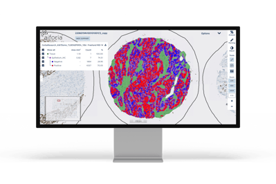
Cerba Research stained 192 tumors of various origins, including breast and prostate, with the CONFIRM anti-Ki-67 clone (30-9) (ROCHE monoclonal primary antibody (IVD)) on the Ventana Benchmark Ultra. Three pathologists were appropriately trained following the International Ki-67 Working Group (IKWG) recommendations and scored tissues accordingly.³
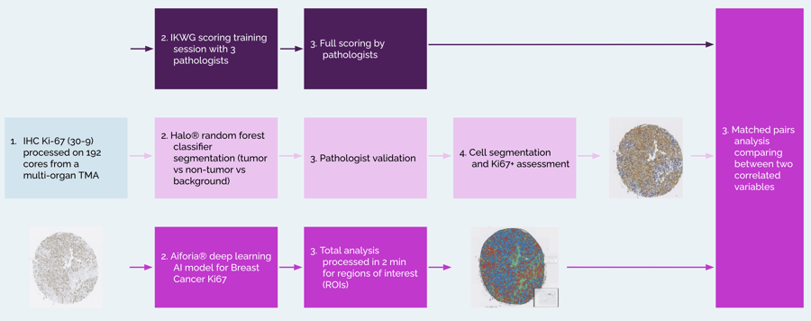
Out of 192 cores, only 158 were analyzed due to the absence of tissue and/or pathologists' inability to score. Depending on the analysis approach applied, Ki-67+ cells were detected in 24.38 - 28.71% of the tumor cells. The study shows a very high consistency of results obtained for Ki-67 scoring between the two image analysis software (r2=0.95) on solid tumors analyzed (n=158). However, the correlation obtained between the pathologists was weaker (mean r2=0.83), despite appropriate training and following guidelines, but remains within an acceptable range.
Rania Gaspo, Global Therapy Area Lead from Cerba Research, comments on the results: “In a nutshell, this work shows that recent AI-based image analysis tools, such as the Aiforia® Platform, provide valuable assistance in the field of image analysis and allowed us to drastically reduce inter-pathologist variability in the Ki-67 scoring of solid tumors. We were also positively surprised with the speed of execution of the Aiforia® Platform, which was able to process regions of interest for all cores in just 2 minutes, saving us a lot of time and effort.”
Read more on the ESMO 2023 poster: “Pichon X, Gaspo R, Iglesias S, Kumar D, Tliba M, Burrer R, Finan A. Evidence for the utility of artificial intelligence (AI) and image analysis in Ki-67 quantification in solid tumors. Presented at: ESMO annual meeting; October 20-24, 2023; Madrid, Spain”.
References
1. Polley, M. et al. (2015, June). An international study to increase concordance in Ki-67 scoring. Mod Pathol., 28(6), 778-786. doi: https://doi.org/10.1038/modpathol.2015.38
2. Nielsen, T. et al. (2021, July 1). Assessment of Ki-67 in Breast Cancer: Updated Recommendations From the International Ki-67 in Breast Cancer Working Group. J Natl Cancer Inst., 113(7), 808-819. https://doi.org/10.1093/jnci/djaa201
3. Welcome to Ki-67-QC calibrator. URL http://www.gpec.ubc.ca:8080/tmadb-0.1/calibrator/index