What is your job and what does your research focus on?
Marika: I’m an MD, currently a trainee in obstetrics and gynecology and a PhD student in the University of Oulu, in the group of Terhi Piltonen. My research focuses on inflammation, polycystic ovarian syndrome (PCOS) and combined contraceptives.
A summary of your project with Aiforia:
I study endometrial leukocytes and proliferation in PCOS with immunohistochemically stained endometrial samples.
What does Aiforia enable you to do that you otherwise could not (or would find difficult) to with traditional or other methods?
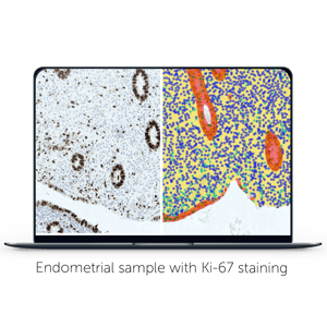 Aiforia enables me to first separate endometrial epithelium and stroma automatically from scanned whole slide images (WSI), which is too difficult a task for traditional image analysis software.
As leukocytes are presented differently in these areas and the amount of epithelium & stroma differs between patients, this separation is crucial for good analysis.
What were your expectations of using deep learning AI?
I hoped that this AI could solve the problem, as otherwise I should either crop my images and rely on older manual techniques or even give up on this valuable project.
Did you know anything about AI before starting your work with Aiforia?
Very little, I had just got a tip that this could be a solution for my problem.
Some details around your project with Aiforia:
Altogether approximately 400 WSI, H-DAB nuclear stainings.
What stage is your algorithm in at the moment?
Algorithm is ready and data is being processed. I’m very happy with the algorithm, it did the job that I wanted it to do!