“With Aiforia, you have the control, you can improve and adapt the methodology…” explains Jenni Säilä, Biomedical Laboratory Scientist at the Helsinki University Hospital.
Working with her supervisor Tiina Vesterinen, a researcher at the HUSLAB Department of Pathology, Jenni developed a deep learning AI model with Aiforia Create to quantify Ki-67 positive and negative tumor cells in rare pulmonary carcinoid (PC) tumors as a part of her thesis study.
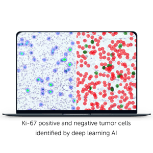
Ki-67 is a commonly used molecular target in cancer diagnostics and its proliferation index is an essential parameter in PC tumor diagnostics.
Traditional methods are fallible
Jenni’s study material consisted of five tissue microarray slides which included altogether 127 PC tumors. The slides were immunohistochemically labelled with a Ki-67 antibody and Jenni used the digitized slides to train her own AI model with Aiforia Create to identify Ki-67 positive and negative tumor cells.
The results Aiforia produced were referenced against a pathologist completing the analysis manually and also compared with results produced by non-AI based image analysis software. The analysis of immunohistochemical stains with these traditional methods is often time consuming and prone to inter- and intra-observer subjectivity.
You don’t have to be perfect to use AI
 ”The AI modeI was better at analyzing the samples. You didn’t have to exclude any areas because Aiforia was able to detect everything. Compared to other image analysis methods in which if there are any issues with the samples like staining errors or broken tissue, the other programs are not able to count in these areas,” describes Jenni.
The convolutional neural networks, which power Aiforia’s deep learning AI, allow image analysis to perform at a level far beyond human capabilities. Perfect training material or high quantities of images or slides are not needed to train or create a robust AI model.
Aiforia is also agile; providing a unique image analysis solution, as AI models can be trained to detect any feature in any image. Tiina explains: ”Traditional image analysis methods you can’t train yourself. You can’t fully customize them to meet your needs. With Aiforia, you have the control, you can improve and adapt the methodology, to find exactly what you are looking for.”
”Our aim is to use this AI algorithm also in clinical diagnostics to assist pathologists. Since preliminary results are promising, we plan to train the algorithm further to calculate Ki-67 proliferation index also for other neuroendocrine tumors,” adds Tiina.